
Casparian strip
The Casparian strip is a band-like thickening in the center of the
.jpg)

The chemistry of the Casparian strip has been described as composed of
Casparian strips differentiate after an outward growth of the
In the absence of secondary growth (most
Discovery
The discovery of the Casparian strip dates back to the mid-19th century, and advances in the understanding of the endodermis of plant roots.[15] In 1865, the German botanist Robert Caspary first described the endodermis of the root of plants, found that its cell wall was thickened, and named it Schuchtzscheide. Later scholars called the thickened part of it the Carls Belt, which was named after Casbury[clarification needed].[5][16] The term "Caspary'schen fleck" (German: Caspary'schen fleck) appeared in the 1870s literature,[17][18] and after the 20th century, it was often called the Casparian strip. In 1922, researchers first left the Casparian strip from the root of plants to study its composition.[clarification needed] [19][20]
Composition
The chemical composition of the Casparian strip has been controversial for a long time. Casbury pointed out that this structure may be composed of lignin or suberin. Later scholars mostly thought it was suberin.[21] It was not until the 1990s that after analyzing the Casparian strip of several plants, it was found that lignin was the main component, but many textbooks have not been updated.[4] Although the cell wall of the endothelium is rich in woodbolic, this is the result of the sublevel differentiation of the endothelium.[note 1] In the past, some scholars believe that the formation of the endodermis of Casparian strip is the beginning of sublevel differentiation, but there is no direct relationship between the two. The casparian strip has formed after the primary differentiation, and the secondary differentiation begins with the slash cut of the root, not where the Casparian strip is.[1]
Function
The casparian strip is fully filled with the gap between endothelial cells, including the middle lamella, making the cell walls of the two cells almost fused.[1] In the transportation of water and inorganic nutrients at the root of plants, the Casparian strip mainly affects the transportation of primary in vitro, that is, the transportation of water and inorganic salts through the interstitial cells of the epidermis and cortex cells. When water and inorganic salt come to the endothelial cells, they need to enter the cell through the cell membrane because the casparian strip is not water-permeable, and then transported by the protoplasmic inner path to reach the lignan cells of the stele, and then to other organs such as the stems and leaves.[16] When the growth environment is unfavourable, the casparian strip can act as a barrier between plant cells and the outside world, avoiding the entry of ions or outflow of their own ions in the environment.[7] In addition, the thickening of the carcass belt and the cortex also prevents toxic substances or pathogen invasion, as well as the function of preventing water dispersion.[22] Some studies have shown that plants may form thicker Casparian strip in high-salt environments, and in areas closer to the tip of the roots, which may be an adaptation to the environment,[23][24] but compared with the endothelial sublevel differentiated wooden bolt walls, which are significantly thickened in high-salt adversity, the Casparian strip changes is smaller.[25]
The Casparian strip is mainly located in the endodermis of the root,[26] but some plants also have the Casparian strip in the outer cortex on the outer side of the root cortex, stem or leaf.[27] For example, the conifers of Pinus bungeana and the stems of Pelargonium have the Casparian strip, which may be related to preventing water dispersion or pathogenic invasion.[28][29]
Development

The development of the Casparian strip is started after the endogenic cells are fully delayed,[21][30] and there is currently two news signal transduction that promote endothelial cell formation of Casparian strip. The first is transcription factor Short-root (SHR) Activated additional two transcription factors Myb36 and Scarecrow (SCR), the former can stimulate Casparian Strip Proteins (Casp1-5), Peroxidase (PER64) and ESB1 (Enhanced) Suberin 1), etc., the latter affects the position of the Casparian strip in the inner skin cell, which causes the position of the Casparian strip to be too close to the Stele;[6] the second is medium Casparian Strip Integrity Factor (CIF1-2) and the GSO1 (SGN3) and GSO2 receptor bonded to the endothelial cell radial wall and the GSO2 receptor in the lateral wall. CASP in the cells is concentrated to a cell membrane region corresponding to the position of the Casparian strip, forming a Casparian Strip Membrane Domain (CSD), and the CSD is incorporated in the region. The GS01 receptor is surrounded by the edge of each CSD region, promoting CSD fused into a continuous strip region, that is, the region where the Casparian strip is to be formed.[7][31]
Casparian strip protein is a membrane protein that interacts with each other and can bind to proteins needed to synthesize lignin such as PER64, ESB1 and respiratory oxidase homologer F (RBOHF) to activate the downstream reaction of Casparian strip development.[1][5] In mutant plants lacking GSO1 receptors or at the same time lacking CIF1 and CIF2 polypeptides, CASP1 is abnormally distributed on the endothelial cell membrane, and the CSD cannot normally fuse into a continuous and complete band structure, thus eventually forming a broken and discontinuous Casparian strip.[7][31]
Environmental factors such as light, soil salinity and water deficit can affect the development of the Casparian strip.[28]
Photo
-
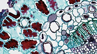
 The vascular bundles of the root of the genusmonocotyledons) can be seen in the endothelium and the Casparian strip around the middle column. The Casparian strip is dyed red because it contains lignin.
 -
 The vascular bundle of the root of the genus Ranunculus (dicotyledon) can see the endothelium and the Casparian strip around the middle column. The Casparian strip is dyed red because it contains lignin.
 -
 A Pinaceae (Gymnosperm) coniferous leaves. Peripheral endothelial endodermis and Casparian strip of visible vascular bundles
.jpg)
.jpg)
.jpg)
See also
Notes
- ^ After the endothelial cell wall in the old root forms a wood embolism thickened, its function can be changed from transmission water to protecting plants, which can further limit the transmission of water and inorganic salts. Only channel cells (a few sublevel differentiated endothelial cells) retain transportation function. As the root grows, some plants lose channel cells in the root.[1]
References
- ^ PMID 23451777.
- PMID 21904117.
- ISBN 978-0-12-409751-3, retrieved 2022-12-22
- ^ a b Geldner, N. (2013). "Casparian strips" (PDF). Current Biology. Vol. 23, no. 23. pp. R1025, R1026.
- ^ S2CID 4366553.
- ^ PMID 30057307.
- ^ S2CID 206653442.
- S2CID 45393531.
- ^ a b Frey-Wyssling, A.; H. H. Bosshard (1959). Cytology of the Ray Cells in Sapwood and Heartwood. Cram.
- ^ a b Taiz, L., Zeiger, Eduardo, Møller, Ian Max, & Murphy, Angus. (2015). Plant physiology and development (Sixth ed.).
- S2CID 873056.
- ^ von Guttenberg, H. (1943). Die physiologischen Scheiden. Borntraeger.
- ^ OGURA, Y. (1938). "Problems in morphology (13)". Botany and Zoology. 6: 139–148.
- ^ Napp-Zinn, A. F. (1953). 100 Jahre Köln-düsseldorfer Rheindampfschiffahrt: Insbesondere Zerstörung und Wiederaufbau 1939-1953. Köln-Düsseldorfer Rheindampfshiffahrt.
- .
- ^ S2CID 873056.
- ^ Russow, E. (1872). "Rhizocarpeae. I. Axenorgane: A. Stamm: a. Marsilia (Drummondii, elata, salvatrix)". Vergleichende Untersuchungen betreffend die Histiologie (Histiographie und Histiogenie) der vegetativen und Sporen-bildenden Organe und die Entwickelung der Sporen der Leitbündel-Kryptogamen: mit Berücksichtigung der Histiologie der Phanerogamen, ausgehend von der Betrachtung der Marsiliaceen. Commissionnaires de l'Académie Impériale des sciences. pp. 1–12.
- ^ Müller, C. (1884). "Morphologie der Gewebe". 12 (1): 234–342.
{{cite journal}}: Cite journal requires|journal=(help) - JSTOR 2428118.)
{{cite journal}}: CS1 maint: multiple names: authors list (link - ISSN 0931-1890.
- ^ PMID 22665765.)
{{cite journal}}: CS1 maint: multiple names: authors list (link - PMID 25125504.)
{{cite journal}}: CS1 maint: multiple names: authors list (link - S2CID 792699.)
{{cite journal}}: CS1 maint: multiple names: authors list (link - PMID 21904117.)
{{cite journal}}: CS1 maint: multiple names: authors list (link - PMID 30517104.
- ISBN 978-0-470-04737-8. Archived from the originalon 2020-12-01. Retrieved 2021-04-17.
- S2CID 45859773.
- ^ PMID 16170202.
- PMID 21239408.)
{{cite journal}}: CS1 maint: multiple names: authors list (link - PMID 27551946.
- ^ PMID 25233277.)
{{cite journal}}: CS1 maint: multiple names: authors list (link
- Esau, Katherine (1965). Plant Anatomy. John Wiley & Sons. p. 767. ISBN 9780471244554.