
Subclavian vein
This article needs additional citations for verification. (July 2019) |
| Subclavian vein | |
|---|---|
upper extremity | |
| Source | axillary vein, external jugular vein |
| Drains to | brachiocephalic vein |
| Artery | subclavian artery |
| Identifiers | |
| Latin | vena subclavia |
| MeSH | D013350 |
| TA98 | A12.3.08.002 |
| TA2 | 4953 |
| FMA | 4725 |
| Anatomical terminology] | |
The subclavian vein is a paired large vein, one on either side of the body, that is responsible for draining blood from the upper extremities, allowing this blood to return to the heart. The left subclavian vein plays a key role in the absorption of lipids, by allowing products that have been carried by lymph in the thoracic duct to enter the bloodstream. The diameter of the subclavian veins is approximately 1–2 cm, depending on the individual.[medical citation needed]
Structure
Each subclavian vein is a continuation of the
The subclavian vein follows the
Function
The
The right lymphatic duct drains its lymph into the junction of the right internal jugular vein, and the right subclavian vein.
Clinical relevance
Central venous lines
As the subclavian vein is large, central and relatively superficial, the right subclavian vein is often used to place
Thoracic outlet syndrome
The subclavian vein may be blocked during thoracic outlet syndrome.[7] This can lead to arm swelling, pain, and cyanosis.[7] The cause of the thoracic outlet syndrome, whether a thrombus or external pressure, must be reversed urgently.[7]
Etymology
Sub (below), and clavian (pertaining to the clavicle).
Disorders
Paget–Schroetter disease includes the thrombosis of the subclavian veins, in this case usually caused by exercise-induced strains.
See also
Additional images
-
 Peculiar ribs.
 -
 The venæ cavæ and azygos veins, with their tributaries.
 -
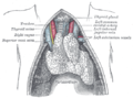
 The thoracic and right lymphatic ducts.
 -
 The thymus of a full-term infant, exposed in situ.
 -
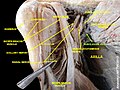
 Subclavian vein
 -
 Subclavian vein - right view
 -
 Subclavian vein
 -
 Subclavian vein


References
- ^ ISBN 978-0-443-10373-5, retrieved 2020-11-20
- ISBN 978-1-4160-6208-0, retrieved 2020-11-20
- ^ "What is the Subclavian Vein? (with pictures)". wiseGEEK. Retrieved 2019-01-03.
- ISBN 978-1-4160-3786-6, retrieved 2020-11-20
- ^ S2CID 81592410, retrieved 2020-11-20
- ISBN 978-0-323-03004-5, retrieved 2020-11-20
- ^ ISBN 978-0-12-369515-4, retrieved 2020-11-20