
Fallopian tube
| Fallopian tube | |
|---|---|
Lumbar lymph nodes | |
| Identifiers | |
| Latin | tuba uterina |
| Greek | σάλπιγξ (sálpinx) |
| MeSH | D005187 |
| TA98 | A09.1.02.001 |
| TA2 | 3486 |
| FMA | 18245 |
| Anatomical terminology] | |
The fallopian tubes, also known as uterine tubes, oviducts[1] or salpinges (sg.: salpinx), are paired tubular sex organs in the human female body that stretch from the ovaries to the uterus. The fallopian tubes are part of the female reproductive system. In other vertebrates, they are only called oviducts.[2]
Each tube is a muscular
An
Almost a third of cases of infertility are caused by fallopian tube pathologies. These include inflammation, and tubal obstructions. A number of tubal pathologies cause damage to the cilia of the tube, which can impede movement of the sperm or egg.[7]
The name comes from the
Structure
Each fallopian tube leaves the uterus at an opening at the
Parts
Each tube is composed of four parts: from inside the proximal tubal opening the intramural or interstitial part, that links to the narrow isthmus, the isthmus connects to the larger ampulla, which connects with the infundibulum and its associated fimbriae that opens into the peritoneal cavity from the distal tubal opening.[12]
Intramural part
The intramural part or interstitial part of the fallopian tube lies in the myometrium, the muscular wall of the uterus. This is the narrowest part of the tube that crosses the uterus wall to connect with the isthmus. The intramural part is 0.7 mm wide and 1 cm long.[12]
Isthmus
The narrow isthmus links the tube to the uterus, and connects to the ampulla. The isthmus is a rounded, and firm muscular part of the tube. The isthmus is 1–5 mm wide, and 3 cm long.[12] The isthmus contains a large number of secretory cells.[10]
Ampulla
The ampulla is the major part of the fallopian tube. The ampulla is the widest part of the tube with a maximal luminal diameter of 1 cm, and a length of 5 cm. It curves over the ovary, and is the primary site of fertilization.[12] The ampulla contains a large number of ciliated epithelial cells.[10] It is thin walled with a much folded luminal surface, and opens into the infundibulum.[12]
Infundibulum
The infundibulum opens into the abdomen at the distal tubal opening and rests above the ovary. Most cells here are ciliated epithelial cells.[10] The opening is surrounded by fimbriae, which help in the collection of the oocyte after ovulation.[4] The fimbriae (singular fimbria) is a fringe of densely ciliated tissue projections of approximately 1 mm in width around the distal tubal opening, oriented towards the ovary.[12] They are attached to the ends of the infundibulum, extending from its inner circumference, and muscular wall.[12] The cilia beat towards the fallopian tube.[12] Of all the fimbriae, one fimbria known as the ovarian fimbria is long enough to reach and make contact with the near part of the ovary during ovulation.[13][14][12] The fimbriae have a higher density of blood vessels than the other parts of the tube, and the ovarian fimbria is seen to have an even higher density.[8]
An ovary is not directly connected to its adjacent fallopian tube. When ovulation is about to occur, the
Microanatomy

When
The outermost covering layer of serous membrane is known as the serosa.[6] The serosa is derived from the visceral peritoneum.[14]
The muscularis mucosae consists of an outer ring of
The innermost mucosa is made up of a layer of luminal epithelium, and an underlying thin layer of
The histological features of tube vary along its length. The mucosa of the ampulla contains an extensive array of complex folds, whereas the relatively narrow isthmus has a thick muscular coat and simple mucosal folds.[14]
Development

As the uterus develops, the part of the fallopian tubes closer to the uterus, the ampulla, becomes larger. Extensions from the fallopian tubes, the fimbriae, develop over time. Cell markers have been identified in the fimbriae, which suggests that their embryonic origin is different from that of the other tube segments.[8]
Apart from the presence of sex chromosomes, specific genes associated with the development of the fallopian tubes include the Wnt and Hox groups of genes, Lim1, Pax2, and Emx2.[17]
Embryos have two pairs of ducts that will let gametes out of the body when they are adults; the paramesonephric ducts develop in females into the fallopian tubes, uterus, and vagina.
Function
Fertilization

The fallopian tube allows the passage of an egg from the ovary to the uterus. When an
At the time of
The release of an oocyte does not alternate between the two ovaries and seems to be random. After removal of an ovary, the remaining one produces an egg every month.[18]
Clinical significance
Almost a third of cases of infertility are caused due to fallopian tube pathologies. These include inflammation, and tubal obstructions. A number of tubal pathologies cause damage to the cilia of the tube, which can impede movement of the sperm or egg. A number of sexually transmitted infections can lead to infertility.[7]
Inflammation
Salpingitis is inflammation of the fallopian tubes and may be found alone, or with other pelvic inflammatory diseases (PIDs). A thickening of the fallopian tube at its narrow isthmus portion, due to inflammation, is known as salpingitis isthmica nodosa. Like another PID endometriosis, it may lead to fallopian tube obstruction. Fallopian tube obstruction may be a cause of infertility or ectopic pregnancy.[19]
Blockage or narrowing
If a
Ectopic pregnancy
Occasionally the embryo implants outside of the uterus, creating an ectopic pregnancy. Most ectopic pregnancies occur in the fallopian tube, and are commonly known as tubal pregnancies.[22]
Surgery

The surgical removal of a fallopian tube is called a salpingectomy. To remove both tubes is a bilateral salpingectomy. An operation that combines the removal of a fallopian tube with the removal of at least one ovary is a salpingo-oophorectomy. An operation to remove a fallopian tube obstruction is called a tuboplasty. A surgical procedure to permanently prevent conception is tubal ligation.
Cancer
Other
In rare cases, a fallopian tube may prolapse into the vaginal canal following a hysterectomy. The swollen fimbriae can have the appearance of an adenocarcinoma.[24]
History
The Greek doctor Herophilus, in his treatise on midwifery, points out the existence of the two ducts that he supposed transported "female semen". Then Galen, already in the modern era, described that the paired ducts indicated by Herophilus were connected to the uterus.
In 1561, the Renaissance doctor Gabriele Falloppio published his book Observationes Anatomicae. Its contribution is a detailed description of the "tubal" of the uterus and its different portions, with its farthest (distal) end open towards the abdomen, and the other (proximal) connected to the uterus.[25][26]
Though the name Fallopian tube is
Additional images
-
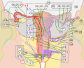
 Image showing numbered parts of the fallopian tubes and surrounding structures
 -
 Female reproductive system numbered parts
 -
 Image showing the right fallopian tube (here labeled the uterine tube) seen from behind. The uterus, ovaries and right broad ligament are labeled.
 -
 Cross-section of fallopian tube, stained and viewed under microscope


References
![]() This article incorporates text in the public domain from page 1257 of the 20th edition of Gray's Anatomy (1918)
This article incorporates text in the public domain from page 1257 of the 20th edition of Gray's Anatomy (1918)
- ^ "Uterine Tube (Fallopian Tube) Anatomy: Overview, Pathophysiological Variants". 14 July 2021. Retrieved 15 September 2022.
- PMID 25399777.
- PMID 31613440. Retrieved 22 September 2022.
- ^ a b c d "Fallopian Tube Disorders: Overview, Salpingitis and Pelvic Inflammatory Disease, Salpingitis Isthmica Nodosa". 15 March 2022. Retrieved 17 September 2022.
- ^ PMID 29763118. Retrieved 25 September 2022.
- ^ ISBN 9780470233474.
- ^ PMID 25866566.
- ^ PMID 32587423.
- PMID 10207477. Archived from the originalon 15 April 2013. Retrieved 28 May 2010.
- ^ S2CID 27164540.
- PMID 33167378.
- ^ ISBN 9780702052309.)
{{cite book}}: CS1 maint: location missing publisher (link - ^ "ovarian fimbria". cancerweb.ncl.ac.uk. Archived from the original on 21 June 2008. Retrieved 2 May 2022.
- ^ ISBN 9788131225561.
- ^ "Dictionary - Normal: Fallopian tube - The Human Protein Atlas". www.proteinatlas.org. Retrieved 25 October 2022.
- ^ PMID 35644062.
- ^ ISBN 9781441904881.
- Merck Manual. Retrieved 6 March 2011.
- ^ Salpingitis at eMedicine
- ^ "Tubal Factor Infertility (Fallopian Tube Obstruction) | ColumbiaDoctors - New York". ColumbiaDoctors. 20 June 2017. Retrieved 30 June 2022.
- S2CID 43312882.
- ^ "Ectopic pregnancy | RCOG". Retrieved 2 October 2022.
- PMID 34669199.
- ISSN 1756-2228. Retrieved 11 March 2018.
- ISSN 0717-9502.
- ISSN 2304-5132.
- ^ "Definition of FALLOPIAN TUBE". www.merriam-webster.com. Retrieved 26 September 2022.
- .
External links
- Histology image: 18501loa – Histology Learning System at Boston University
- Menstrual Cycle - Merck