
Levator palpebrae superioris muscle
| Levator palpebrae superioris | ||
|---|---|---|
Antagonist Palpebral part of orbicularis oculi muscle | | |
| Identifiers | ||
| Latin | musculus levator palpebrae superioris | |
| TA98 | A15.2.07.020 | |
| TA2 | 2052 | |
| FMA | 49041 | |
| Anatomical terms of muscle] | ||
The levator palpebrae superioris (
Structure
The levator palpebrae superioris originates from inferior surface of the lesser wing of the
Blood supply
The levator palebrae superioris receives its blood supply from branches of the ophthalmic artery, specifically, muscular branches and the supraorbital artery. Blood is drained into the superior ophthalmic vein.
Nerve supply
The levator palpebrae superioris receives motor innervation from the superior division of the oculomotor nerve.[1][2][3] The smooth muscle that originates from its undersurface, called the superior tarsal muscle is innervated by postganglionic sympathetic axons from the superior cervical ganglion.[2]
Function
The levator palpebrae superioris elevates the upper eyelid.[1][2]
Clinical significance
Damage to this muscle or its innervation can cause ptosis, which is drooping of the eyelid.[4][5] Lesions in CN III can cause ptosis,[5] because without stimulation from the oculomotor nerve the levator palpebrae cannot oppose the force of gravity, and the eyelid droops.
Ptosis can also result from damage to the adjoining superior tarsal muscle or its sympathetic innervation. Such damage to the sympathetic supply occurs in Horner's syndrome and presents as a partial ptosis. It is important to distinguish between these two very different causes of ptosis. This can usually be done clinically without issue, as each type of ptosis is accompanied by other distinct clinical findings.
The ptosis seen in paralysis of the levator palpebrae superioris is usually more pronounced than that seen due to paralysis of the superior tarsal muscle.
Additional images
-
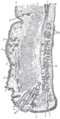 Sagittal section through the upper eyelid.
 -
 Levator palpebrae superioris muscle
 -
 Levator palpebrae superioris muscle
 -
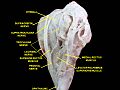 Extrinsic eye muscle. Nerves of orbita. Deep dissection.
 -
 Extrinsic eye muscle. Nerves of orbita. Deep dissection.


See also
References
- ^ ISBN 978-0-443-06557-6, retrieved 2020-11-11
- ^ )
- S2CID 241607885, retrieved 2020-11-11
- ISBN 978-0-323-04456-1
- ^ ISBN 978-0-12-802381-5, retrieved 2020-11-11
External links
- Anatomy figure: 29:01-01 at Human Anatomy Online, SUNY Downstate Medical Center
- lesson3 at The Anatomy Lesson by Wesley Norman (Georgetown University) (orbit2)