
Scanning electrochemical microscopy
Scanning electrochemical microscopy (SECM) is a technique within the broader class of
The technique is complementary to other surface characterization methods such as surface plasmon resonance (SPR),[8]
History
The emergence of
Principles of operation
Electric potential is manipulated through the UME tip in a bulk solution containing a redox-active couple (e.g. Fe2+/Fe3+). When a sufficiently negative potential is applied, (Fe3+) is reduced to (Fe2+) at the UME tip, generating a diffusion-limited current.[13] The steady-state current is governed by the flux of oxidized species in solution to the UME disc and is given by:

where iT,∞ is the diffusion-limited current, n is the number of electrons transferred at the electrode tip (O + ne− → R), F is
There are two predominant modes of operation, which are feedback mode and collection-generation mode.
Feedback mode

In a bulk solution, the oxidized species is reduced at the tip, producing a steady-state current that is limited by hemispherical diffusion. As the tip approaches a conductive substrate in the solution, the reduced species formed at the tip is oxidized at the conductive surface, yielding an increase in the tip current and creating a regenerative "positive" feedback loop.[6] The opposite effect is observed when probing insulating surfaces, as the oxidized species cannot be regenerated and diffusion to the electrode is inhibited as a result of physical obstruction as the tip approaches the substrate, creating a "negative" feedback loop and decreasing the tip current. An additional parameter to consider when probing insulating surfaces is the electrode sheath diameter, rg, since it contributes to the physical obstruction of diffusion.
The change in tip current as a function of distance d can be plotted as an "approach curve" as shown.
Due to the rate dependent nature of SECM measurements, it is also employed to study electron-transfer kinetics.[17]
Collection-generation modes
Another mode of operation that is employed is tip generation/substrate collection (TG/SC). In TG/SC mode, the tip is held at a potential sufficient for an electrode reaction to occur and "generate" a product while the substrate is held at a potential sufficient for the electrode product to react with or be "collected" by the substrate.[6] The reciprocal to this method is substrate generation/tip collection (SG/TC), where the substrate acts to generate a species that is measured at the tip. Both TG/SC and SG/TC variations are also categorized as "direct" modes.[7]
Two currents are generated: the tip current, iT, and the substrate current, iS. Since the substrate is generally much larger than the tip, the efficiency of collection, iS/iT, is 1 if no reactions occur during the transfer of tip-generated species to the substrate. As the distance between tip and substrate, d, decreases, the collection efficiency, iS/iT, approaches 1.
Alternating Current (ac)-SECM
In ac-SECM a sinusoidal bias is applied to the dc bias of the SECM probe allowing the impedance of a sample to be measured, as is the case in electrochemical impedance spectroscopy.[18] Unlike dc-SECM techniques ac-SECM does not require the use of a redox mediator. This is particularly advantageous for measurements where the redox mediator could affect the chemistry of the system under study.[19] Examples include corrosion studies where a redox mediator may act to inhibit or enhance the rate of corrosion, and biological studies where a redox mediator may be toxic to the living cell under study.
In ac-SECM the feedback response measured is dependent on both the sample type and the experimental conditions.[20] When a sample is insulating the measured impedance will always increase with decreasing probe to sample distance. This is not the case for a conductive sample however. For a conductive sample measured in a high conductivity electrolyte, or measured with a low ac frequency, decreasing the probe to sample distance will lead to an increase in impedance. If, however, a conductive sample is measured in a low conductivity electrolyte, or with a high ac frequency, decreasing the probe to sample distance will result in a lower measured impedance.

SECM imaging
Changes in current as a function of distance between electrode tip and substrate surface allow imaging of insulating and conducting surfaces for topology and reactivity information by moving the tip across surfaces and measuring tip current.
The most common scanning mode is constant-height mode,[7] where the tip height is unchanging and is scanned across the surface in the x-y plane. Alternatively, constant distance measurements are possible, which change the z position to maintain the probe to sample distance as the probe is scanned across the surface in the x-y plane. The constant distance measurement can be based on an electrical signal as is the case in the constant-current mode,[7] where the device attempts to maintain a constant current by changing the substrate to tip distance, d, and recording the change in d. A mechanical signal can also be used to control the probe to sample distance. Examples of this are the intermittent contact (ic)-SECM[21] and shear force[22] techniques which use changes in probe vibration to maintain the probe to sample distance.
Spatial resolution is dependent on the tip radius, the substrate to tip distance, the precision of the electronics, and other considerations.
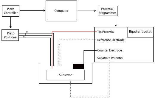
Instrumentation
Early SECMs were constructed solely by individual lab groups from a set of common components including
Preparation of electrodes

SECM probes use platinum as the active core material, however carbon, gold, mercury, and silver have all been used. electropolymerized phenol, and
Modification of electrodes has developed beyond the size parameter. SECM-AFM probes can act as both a force sensor and electrode through the utilization of a flattened, etched metal wire coated by electrophoretic paint. In this system, the flattened wire acts as a flexible cantilever to measure the force against a sample (AFM) as the wire electrode measures the current (SECM).[2] Similarly, SECM functionality can be imparted into standard AFM probes by sputtering the surface with a conductive metal or by milling an insulated tip with a focused ion beam (FIB). Electron-beam lithography has also been demonstrated to reproducibly generate SECM-AFM probes using silicon wafers.[29] AFM probe manufacturers, such as Scuba Probe Technologies fabricate SECM-AFM probes with reliable electrical contacts for operation in liquids.[30]
Images of the chemical environment that is decoupled from localized topographies are also desirable to study larger or uneven surfaces. "Soft stylus probes" were recently developed by filling a microfabricated track on a polyethylene terephthalate sheet with a conductive carbon ink. Lamination with a polymer film produced v-shaped stylus that was cut to expose the carbon tip. The flexibility inherent in the probe design allows for constant contact with the substrate that bends the probe. When dragged across a sample, probe bending accommodates for topographical differences in the substrate and provides a quasi-constant tip-to-substrate distance, d.[31]
Micro-ITIES probes represent another type of specialty probe that utilizes the Interface between Two Immiscible Electrolyte Solutions (ITIES). These tips feature a tapered pipette containing a solution containing a metal counter electrode, and are used to measure electron and ion transfer events when immersed in a second, immiscible liquid phase containing a counter-reference electrode.[1]
Often the probing of liquid/liquid and air/liquid interfaces via SECM require the use of a submarine electrode.[32] In this configuration, the electrode is fashioned into a hook shape where the electrode can be inverted and submerged within the liquid layer. The UME tip points upwards and can be positioned directly beneath the liquid/liquid or air/liquid interface. The portion of the electrode passing through the interface region is electrically insulated to prevent indirect interfacial perturbations.
Increases in the complexity of electrodes along with decreases in size have prompted the need for high resolution characterization techniques.
Potentiostat
The potentiostat biases and measures the voltage using the standard three electrode system of voltammetry experiments. The UME acts as the working electrode to apply a controlled potential to the substrate. The auxiliary electrode (or counter electrode) acts to balance the current generated at the working electrode, often through a redox reaction with the solvent or supporting electrolyte. Voltage measured with regard to the well defined reduction potential of the reference electrode, although this electrode itself does not pass any current.
Positioners and translators
SECM utilizes many of the same positioning components that are available to other materials characterization techniques. Precise positioning between the tip and sample is an important factor that is complementary to tip size. The position of the probe relative to a given point on the material surface in the x, y, and z directions is typically controlled by a motor for rough positioning coupled with a piezoelectric motor for finer control. More specifically, systems may feature an inchworm motor that directs coarse positioning with additional z control governed by a PZT piezo pusher. Stepper motors with XYZ piezo block positioner or closed-loop controller systems have also been used.[15]
Applications
SECM has been employed to probe the topography and surface reactivity of solid-state materials, track the dissolution kinetics of ionic crystals in aqueous environments, screen electrocatalytic prospects, elucidate enzymatic activities, and investigate dynamic transport across synthetic/natural membranes and other biophysical systems. Early experiments focused on these solid/liquid interfaces and the characterization of typical solution-based electrochemical systems at higher spatial resolution and sensitivities than bulk electrochemical experiments typically afford. More recently the SECM technique has been adapted to explore the chemical transfer dynamics at liquid/liquid and liquid/gas interfaces.
Solid/Liquid Interface
Microstructuring
SECM and variations of the technique have also found use in microfabrication, surface patterning, and microstructuring.
Varieties of SECM employing the micropipet tip geometry have been used to generate spatially resolved microcrystals of a
Ionic dissolution

The
Early examples demonstrating the utility of SECM to extract quantitative rate data from such systems was carried out on CuSO4 crystals in an aqueous solution saturated with Cu2+ and SO2−
4 ions.
Electrocatalysis investigation
Approaching the search for novel catalytic materials to replace precious metals used in
A variety of approaches have been suggested for high throughput assessment of novel metallic electrocatalysts. One functional, non-SECM approach, enabled the electrocatalytic activities of a large number of catalysts to be assessed optically by employing a technique that detected
Biological analysis

The ability to probe non-conductive surfaces makes SECM a feasible method for analyzing membranes, redox active enzymes, and other biophysical systems.
Changes in intracellular redox activity may be related to conditions such as
Transport of ions such as K+ and Na+ across membranes or other biological interfaces is vital to many cell processes; SECM has been employed in studying transport of redox active species across cell membranes. In feedback mode, the transfer of molecules across a membrane can be induced by collecting the transferred species at the tip and forming a concentration gradient.[4] The changes in current can be measured as a function of molecule transport rate.
Liquid/liquid interface
Electrocatalysis
The interface between two immiscible electrolyte solutions (ITIES) can be studied using SECM with a micro-ITIES probe. The probe lies in one layer, and is moved closer to the junction while applying a potential. Oxidation or reduction depletes the substrate concentration, resulting in diffusion from either layer. At close tip-interface distances, rates of diffusion between the organic/aqueous layer for a substrate or ionic species are observed.[43] Electron transfer rates have also been studied extensively at the ITIES. In such experiments, redox couples are dissolved in separate phases and the current at the ITIES is recorded.[1] This is also the fundamental principle in studying transport across membranes.
Liquid/gas interface
The transfer of chemical species across air/liquid interfaces is integral to almost every physical, physiological, biological and environmental system on some level. Thus far, a major thrust in the field has been the quantification of molecular transfer dynamics across
Though much work has been done in the area of
References
- ^ a b c Unwin, Patrick; Barker, Gonsalves; Macpherson, Slevin (1999). "Scanning electrochemical microscopy: beyond the solid/liquid interface". 385: 223–240.
{{cite journal}}: Cite journal requires|journal=(help) - ^ .
- PMID 16970330.
- ^ PMID 17287874.
- ISBN 978-3-540-42583-0.
- ^ ISSN 0003-2700.
- ^ ISBN 0-8247-0471-1.
- PMID 15461512.
- .
- PMID 15231302.
- PMID 17285666.
- ^ PMID 14611266.
- ^ .
- .
- ^ PMID 17287874. Retrieved 5 October 2011.
- PMID 22031463.
- .
- ^ "Introduction to ac-SECM" (PDF). Bio-Logic Science Instruments. Retrieved 2019-05-29.
- PMID 8311247.
- ISSN 0013-4686.
- PMID 20583818.
- PMID 12740850.
- .
- S2CID 18506777. Retrieved 5 October 2011.
- PMID 27736057.
- ^ P. Sun, Z. Zhang, J. Guo and Y. Shao, Anal. Chem., 2001, 73, 5346.
- ^ C. J. Slevin, N. J. Gray, J. V. Macpherson, M. A. Webb and P. R. Unwin, Electrochem. Commun., 1999, 1, 282.
- ^ Amemiya S, Bard AJ, Fan FR, Mirkin MV, Unwin PR. Annu Rev Anal Chem (Palo Alto Calif). 2008;1:95-131.
- ^ Dobson P S, Weaver J M R, Holder M N, Unwin P R and Macpherson J V 2005 Characterization of batch microfabricated scanning electrochemical–atomic force microscopy probes Anal. Chem. 77 424–34
- ^ "Electrochemical Cantilevers | Scuba Probe Technologies". 7 September 2017.
- ^ Fernando Cortés-Salazar, Markus Träuble, Fei Li, Jean-Marc Busnel, Anne-Laure Gassner, Mohamad Hojeij, Gunther Wittstock, Hubert H Girault. "Soft Stylus Probes for Scanning Electrochemical Microscopy" Analytical Chemistry Vol. 18, Issue 16. Date: 08/15/2009 Start Page: 6889.
- .
- PMID 10952523.
- PMID 21793140.
- .
- .
- .
- ^ PMID 15631486.
- PMID 12964739.
- PMID 9624047.
- PMID 10963658.
- .
- .
- ISBN 9780471612957.)
{{cite book}}: CS1 maint: location missing publisher (link - .
| Common |
|  |
|---|---|---|
| Other |
| |
| Applications | ||
| See also | ||