
Anterior chamber of eyeball
| Anterior chamber of eyeball | |
|---|---|
 Anterior part of human eye, with anterior chamber at right. | |
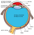
 Schematic diagram of the human eye. | |
| Details | |
| Identifiers | |
| Latin | camera anterior bulbi oculi |
| Acronym(s) | AC |
| MeSH | D000867 |
| TA98 | A15.2.06.003 |
| TA2 | 6792 |
| FMA | 58078 |
| Anatomical terminology | |
The anterior chamber (
The depth of the anterior chamber of the eye varies between 1.5 and 4.0 mm, averaging 3.0 mm. It tends to become shallower at older age and in eyes with
Clinical significance
Depth measurement
Determining the anterior chamber depth (ACD) is important in estimating the risk of angle closure glaucoma. There are various method of measuring ACD, including examination through a slit lamp, ultrasound and Scheimpflug photography. These methods require sophisticated examination equipment and expertise.
A simpler clinical method of quantitatively estimating ACD using smartphone photography (EZ ratio) was developed by Dr Ehud Zamir from the Centre for Eye Research Australia, the University of Melbourne, and published in 2016.[2]
EZ ratio method
The EZ ratio method is one way to calculate the estimated anterior chamber depth.[2] To start, the patient looks at a target in the distance with one eye covered. The examiner takes a digital photograph of the open, examined eye, from the side, perpendicular to the visual axis (a profile photograph).
The following parameters then need to be measured in the photograph, using a personal computer or a smartphone (figures 1,2):
1. The pixel distance between the limbus (the junction between clear cornea and white sclera) and the front of the cornea. This distance is referred to as Z.
2. The pixel distance between the limbus and the centre of the pupil. This distance is referred to as E.
E:Z ratio is the arithmetic ratio between E and Z.
This
Anterior chamber depth (expressed in millimetres) = -3.3 x EZ ratio + 4.2
This estimate has been shown to be accurate with a 95% confidence interval of +/– 0.33 mm error, when compared to measurements of the anterior chamber depth by Scheimpflug photography.[2]


Associated immune deviation
One peculiar feature of the anterior chamber is dampened immune response to allogenic grafts. This is called anterior chamber associated immune deviation (ACAID), a term introduced in 1981 by Streilein et al.[3][4] This phenomenon is relevant to the fact that the eye is considered an "immune privileged site", like the brain and the testis.
Pathology
Additional images
-
 Anterior chamber angle cross-section imaged by anSD-OCT.
 -
 Gonioscopy of the anterior chamber angle
 -
 Gonioscopy of the anterior chamber angle. Labeled structures: 1. Schwalbe's line, 2. Trabecular meshwork (TM), 3. Scleral spur, 4. Ciliary body, 5. Iris
 -
 Anterior chamber IOL
 -
 Structures of the eye labeled
 -
 This image shows another labeled view of the structures of the eye
.png)

.jpg)


See also
- Anterior segment
- Anterior chamber IOL (phakic IOL)
- Anterior chamber paracentesis
References
- ISBN 978-0-937404-33-1.
- ^ PMID 27540496.
- PMID 6788883.
- ^ "Research Story - sce.com". Archived from the original on 2015-02-11. Retrieved 2012-07-16.
External links
- Atlas image: eye_2 at the University of Michigan Health System - "Sagittal Section Through the Eyeball"
| Fibrous tunic (outer) |
|  | |||||
|---|---|---|---|---|---|---|---|
| Uvea / vascular tunic (middle) |
| ||||||
| Retina (inner) |
| ||||||
| Anatomical regions of the eye |
| ||||||
| Other |
| ||||||
| National | |
|---|---|
| Other | |