
Iris (anatomy)
| Iris | |
|---|---|
 | |
 Schematic diagram of the human eye (iris labeled at upper right) | |
| Details | |
| Precursor | Mesoderm and neural ectoderm |
| Part of | Front of eye |
| System | Visual system |
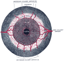
| Artery | long posterior ciliary arteries |
| Nerve | long ciliary nerves, short ciliary nerves |
| Identifiers | |
| Latin | iris |
| MeSH | D007498 |
| TA98 | A15.2.03.020 |
| TA2 | 6753 |
| FMA | 58235 |
| Anatomical terminology | |
The iris (pl.: irides or irises) is a thin, annular structure in the eye in most mammals and birds, responsible for controlling the diameter and size of the pupil, and thus the amount of light reaching the retina. In optical terms, the pupil is the eye's aperture, while the iris is the diaphragm. Eye color is defined by the iris.
Etymology
The word "iris" is derived from the Greek word for "rainbow", also its goddess plus messenger of the gods in the Iliad,[1] because of the many colours of this eye part.[2]
Structure
The iris consists of two layers: the front pigmented fibrovascular layer known as a stroma and, beneath the stroma, pigmented epithelial cells.
The stroma is connected to a

The sphincter pupillae is the opposing muscle of the dilator pupillae. The pupil's diameter, and thus the inner border of the iris, changes size when constricting or dilating. The outer border of the iris does not change size. The constricting muscle is located on the inner border.
The back surface is covered by a heavily pigmented epithelial layer that is two cells thick (the iris pigment epithelium), but the front surface has no epithelium. This anterior surface projects as the dilator muscles. The high pigment content blocks light from passing through the iris to the retina, restricting it to the pupil.[3] The outer edge of the iris, known as the root, is attached to the sclera and the anterior ciliary body. The iris and ciliary body together are known as the anterior uvea. Just in front of the root of the iris is the region referred to as the trabecular meshwork, through which the aqueous humour constantly drains out of the eye, with the result that diseases of the iris often have important effects on intraocular pressure and indirectly on vision. The iris along with the anterior ciliary body provide a secondary pathway for aqueous humour to drain from the eye.

The iris is divided into two major regions:
- The pupillary zone is the inner region whose edge forms the boundary of the pupil.
- The ciliary zone is the rest of the iris that extends to its origin at the ciliary body.
The collarette is the thickest region of the iris, separating the pupillary portion from the ciliary portion. The collarette is a vestige of the coating of the embryonic pupil.[3] It is typically defined as the region where the sphincter muscle and dilator muscle overlap. Radial ridges extend from the periphery to the pupillary zone, to supply the iris with blood vessels. The root of the iris is the thinnest and most peripheral.[4]
The muscle cells of the iris are
Front
- The crypts of Fuchs are a series of openings located on either side of the collarette that allow the stroma and deeper iris tissues to be bathed in aqueous humor. Collagen trabeculae that surround the border of the crypts can be seen in blue irises.
- The midway between the collarette and the origin of the iris: These folds result from changes in the surface of the iris as it dilates.[citation needed]
- Crypts on the base of the iris are additional openings that can be observed close to the outermost part of the ciliary portion of the iris.[4]
Back
- The radial contraction folds of Schwalbe are a series of very fine radial folds in the pupillary portion of the iris extending from the pupillary margin to the collarette. They are associated with the scalloped appearance of the pupillary ruff.
- The structural folds of Schwalbe are radial folds extending from the border of the ciliary and pupillary zones that are much broader and more widely spaced, continuous with the "valleys" between the ciliary processes.
- Some of the circular contraction folds are a fine series of ridges that run near the pupillary margin and vary in thickness of the iris pigment epithelium; others are in ciliary portion of iris.[4]
Microanatomy


From anterior (front) to posterior (back), the layers of the iris are:
- Anterior limiting layer
- Stroma of iris
- Iris sphincter muscle
- Iris dilator muscle (myoepithelium)
- Anterior pigment epithelium
- Posterior pigment epithelium
Development
The stroma and the anterior border layer of the iris are derived from the neural crest, and behind the stroma of the iris, the sphincter pupillae and dilator pupillae muscles, as well as the iris epithelium, develop from optic cup neuroectoderm.
Function
The iris controls the size of the pupil by means of contracting the iris sphincter muscle and/or the iris dilator muscle. The size of the pupils is dependent on many factors (including light, emotional state, cognitive load, arousal, stimulation), and can range from less than 2 mm in diameter, to as large as 9 mm in diameter. However, there is considerable variation in maximal pupil diameter by individual humans, and decreases with age.[6][7] The irises also contract the pupils when accommodation is initiated, to increase the depth of field.
Very few humans possess the ability to exert direct voluntary control over their iris muscles, which grants them the ability to dilate and constrict their pupils on command.[8] However, there is no clear purpose or advantage to this.
Eye color
This article about biology may be excessively human-centric. |

The iris is usually strongly pigmented, with the color typically ranging between brown, hazel, green, gray, and blue. Occasionally, the color of the iris is due to a lack of pigmentation, as in the pinkish-white of oculocutaneous albinism,[3] or to obscuration of its pigment by blood vessels, as in the red of an abnormally vascularised iris. Despite the wide range of colors, the only pigment that contributes substantially to normal iris color is the dark pigment melanin. The quantity of melanin pigment in the iris is one factor in determining the phenotypic eye color of an organism. Structurally, this huge molecule is only slightly different from its equivalent found in skin and hair. Iris color is due to variable amounts of eumelanin (brown/black melanins) and pheomelanin (red/yellow melanins) produced by melanocytes. More of the former is found in brown-eyed people and of the latter in blue- and green-eyed people. The limbal ring appears as a dark ring encircling the iris on some individuals, but is a result of the optical properties of the region between the cornea and sclera, not of pigments in the iris.
Genetic and physical factors determining iris color

Iris color is a highly complex phenomenon consisting of the combined effects of texture, pigmentation, fibrous tissue, and blood vessels within the iris
Melanin is yellowish to dark hazel in the stromal pigment cells, and black in the
The optical mechanisms by which the nonpigmented stromal components influence eye color are complex, and many erroneous statements exist in the literature. Simple selective absorption and reflection by biological molecules (
All the contributing factors towards eye color and its variation are not fully understood. Autosomal recessive/dominant traits in iris color are inherent in other species, but coloration can follow a different pattern.
Different colors in the two eyes
Heterochromia (also known as a heterochromia iridis or heterochromia iridum) is an ocular condition in which one iris is a different color from the other iris (complete heterochromia), or where the part of one iris is a different color from the remainder (partial heterochromia or sectoral heterochromia). Uncommon in humans, it is often an indicator of ocular disease, such as chronic iritis or diffuse iris melanoma, but may also occur as a normal variant. Sectors or patches of strikingly different colors in the same iris are less common.
In contrast, heterochromia and variegated iris patterns are common in veterinary practice.
One eye with a white or bluish-white iris is also known as a "walleye".[13]
Clinical significance
- Angle closure glaucoma
- Aniridia
- Anisocoria
- Horner's syndrome
- Iridocyclitis
- Iridoplegia
- Iritis
- Miosis/Mydriasis
- Synechia
- Third nerve palsy
Alternative medicine
Iridology
Iridology (also known as iridodiagnosis) is an alternative medicine technique whose proponents believe that patterns, colors, and other characteristics of the iris can be examined to determine information about a patient's systemic health. Practitioners match their observations to "iris charts", which divide the iris into zones corresponding to specific parts of the human body. Iridologists see the eyes as "windows" into the body's state of health.[14]
Iridology is not supported by quality research studies,[15] and is considered pseudoscience.[16]
Graphics
-
 Iris, front view
 -
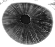 Fluorescein angiograpy of the iris reveals a radial layout of blood vessels.


See also
References
- ^ Liddell, Henry George; Scott, Robert (1940). "ἶρις". A Greek-English Lexicon. Perseus Digital Library.
- ^ "iris". Oxford English Dictionary (Online ed.). Oxford University Press. (Subscription or participating institution membership required.)
- ^ Encyclopædia Britannica 2006 Ultimate Reference Suite DVD
- ^ a b c d e Gold, Daniel H; Lewis, Richard; "Clinical Eye Atlas," pp. 396–397
- ISBN 0-03-910284-X.
- ^ "Aging Eyes and Pupil Size". Amateurastronomy.org. Archived from the original on 2013-10-23. Retrieved 2013-08-28.
- PMID 8125724. Retrieved 2013-08-28.
- ISSN 0167-8760.
- ^ "Sensory Reception: Human Vision: Structure and function of the Human Eye" vol. 27, p. 175 Encyclopædia Britannica, 1987
- JSTOR 44170668.
- S2CID 26240821.
- ^ a b Fabricius, Karl. "Heterochromia in Animals". Environmental Graffiti. Archived from the original on 2010-09-23. Retrieved 2010-10-27.
- ^ "walleye", def. 1a, Merriam-Webster Dictionary
- ^ Novella, Steven. "Iridology". Science-Based Medicine. Archived from the original on 1 July 2017. Retrieved 20 August 2017.
- PMID 10636425.
- ^ Stephen Barrett (9 November 2015). "Iridology Is Nonsense". Quackwatch. Retrieved 6 August 2023.
External links
- Detailed photographs of human irides
- Histology image: 08010loa – Histology Learning System at Boston University
- Atlas image: eye_1 at the University of Michigan Health System – "Sagittal Section Through the Eyeball"
| Fibrous tunic (outer) |
|  | |||||
|---|---|---|---|---|---|---|---|
| Uvea / vascular tunic (middle) |
| ||||||
| Retina (inner) |
| ||||||
| Anatomical regions of the eye |
| ||||||
| Other |
| ||||||