
Desquamation
| Desquamation | |
|---|---|
| Other names | Skin peeling |
.jpg) | |
| Specialty | Dermatology |
Desquamation occurs when the outermost layer of a tissue, such as the
'.Physiologic desquamation
Keratinocytes are the predominant cells of the epidermis, the outermost layer of the skin. Living keratinocytes reside in the basal, spinous, or granular layers of the epidermis. The outermost layer of the epidermis is called the stratum corneum and it is composed of terminally differentiated keratinocytes, the corneocytes. In the absence of disease, desquamation occurs when corneocytes are individually shed unnoticeably from the surface of the skin.[1] Typically the time it takes for a corneocyte to be formed and then shed is about 14 weeks but this time can vary depending on the anatomical location that the skin is covering. For example, desquamation occurs more slowly at acral (palm and sole) surfaces and more rapidly where the skin is thin, such as the eyelids. Normal desquamation can be visualized by immersing skin in warm or hot water. This induces the outermost layer of corneocytes to shed, such as is the case after a hot shower or bath.[citation needed]
Corneocytes are held together by corneodesmosomes. In order for desquamation to occur these corneodesmosome connections must be degraded.
Abnormal desquamation
Scale forms on the skin surface in various disease settings, and is the result of abnormal desquamation. In pathologic desquamation, such as that seen in X-linked ichthyosis, the stratum corneum becomes thicker (hyperkeratosis), imparting a "dry" or scaly appearance to the skin, and instead of detaching as single cells, corneocytes are shed in clusters, which forms visible scales.
-
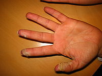
 Desquamation of skin on hands, caused by scarlet fever infection
 -
 Desquamation of skin on fingertips, caused by scarlet fever
 -
 Desquamation of skin on the finger, caused by the popping of an acute paronychia


Eyes
Certain eye tissues, including the
See also
- Desquamative gingivitis
- Exfoliation joint
- Moist desquamation
- Pityriasis—flaking of the skin
- Spalling
- Sunburn
References
- ^ PMID 8460510.
- ^ PMID 35900871.
- PMID 10627489.
- ^ Parillo, Steven J; Parillo, Catherine V. (2010-05-25). "Stevens-Johnson Syndrome". eMedicine. Medcape. Retrieved 2010-09-06.
- ^ Centers for Disease Control and Prevention (2005-06-30). "Cutaneous Radiation Injury". CDC. Retrieved 2011-05-15.
- ^ Gilbard, Jeffrey P. (November 1, 2003). "Dry Eye: Natural History, Diagnosis and Treatment". Wolters Kluwer Pharma Solutions. Archived from the original on January 30, 2013. Retrieved February 3, 2012.
- PMID 18231610.
External links
| Authority control databases: National |
|---|