
Fish scale
A fish scale is a small rigid plate that grows out of the
Scales vary enormously in size, shape, structure, and extent, ranging from strong and rigid armour plates in fishes such as
Fish scales are part of the fish's integumentary system, and are produced from the mesoderm layer of the dermis, which distinguishes them from reptile scales.[2][3] The same genes involved in tooth and hair development in mammals are also involved in scale development. The placoid scales of cartilaginous fishes are also called dermal denticles and are structurally homologous with vertebrate teeth. Most fish are also covered in a layer of mucus or slime which can protect against pathogens such as bacteria, fungi, and viruses, and reduce surface resistance when the fish swims.
Thelodont scales

The bony scales of
Bone, a tissue that is both resistant to mechanical damage and relatively prone to fossilization, often preserves internal detail, which allows the
However, using scale morphology alone to distinguish species has some pitfalls. Within each organism, scale shape varies hugely according to body area,[8] with intermediate forms appearing between different areas—and to make matters worse, scale morphology may not even be constant within one area. To confuse things further, scale morphologies are not unique to taxa, and may be indistinguishable on the same area of two different species.[9]
The morphology and histology of thelodonts provides the main tool for quantifying their diversity and distinguishing between species, although ultimately using such convergent traits is prone to errors. Nonetheless, a framework comprising three groups has been proposed based upon scale morphology and histology.[7] Comparisons to modern shark species have shown that thelodont scales were functionally similar to those of modern cartilaginous fish, and likewise has allowed an extensive comparison between ecological niches.[10]
Cosmoid scales

Cosmoid scales are found only on ancient
Elasmoid scales
Elasmoid scales are thin,
The zebrafish elasmoid scales are used in the lab to study bone mineralization process, and can be cultured (kept) outside of the organism.[13][14]
Ganoid scales
_(3149758934).jpg)

Ganoid scales are found in the
Ganoine is a characteristic component of ganoid scales. It is a glassy, often multi-layered mineralized
 Amblypterus striatus
|
Ganoid scales of the extinct Carboniferous fish, Amblypterus striatus. (a) shows the outer surface of four of the scales, and (b) shows the inner surface of two of the scales. Each of the rhomboidal-shaped ganoid scales of Amblypterus has a ridge on the inner surface which is produced at one end into a projecting peg which fits into a notch in the next scale, similar to the manner in which tiles are pegged together on the roof of a house. | 
|
Most ganoid scales are
-
![The alligator gar has a tough armouring of rhomboidal-shaped ganoid scales.[19]](//upload.wikimedia.org/wikipedia/commons/thumb/b/bd/Alligator_gar_fish.jpg/395px-Alligator_gar_fish.jpg) Therhomboidal-shaped ganoid scales.[19]
Therhomboidal-shaped ganoid scales.[19] -
 The sturgeon has rows of ganoid scales enlarged into scute-like armour plates.
 -
 The ganoid scales on acycloid scales.
![The alligator gar has a tough armouring of rhomboidal-shaped ganoid scales.[19]](/File:Alligator_gar_fish.jpg)
.png)


In sturgeons, the scales are greatly enlarged into armour plates along the sides and back, while in the bowfin the scales are greatly reduced in thickness to resemble cycloid scales.
-
 Earrings made from the ganoid scales of an alligator gar
 -
 Fossil of a primitive rayfin with ganoid scales
 -
 Ganoid scales on a fossilisedmya


.jpg)
Native Americans and people of the Caribbean used the tough ganoid scales of the alligator gar for arrow heads, breastplates, and as shielding to cover plows. In current times jewellery is made from these scales.[20]
Leptoid scales
Leptoid (bony-ridge) scales are found on higher-order bony fish, the
Leptoid scales overlap in a head-to-tail configuration, like roof tiles, making them more flexible than cosmoid and ganoid scales. This arrangement allows a smoother flow of water over the body, and reduces
Leptoid scales come in two forms: cycloid (smooth) and ctenoid (comb-like).[23]
Cycloid scales
Cycloid (circular) scales have a smooth texture and are uniform, with a smooth outer edge or margin. They are most common on fish with soft fin rays, such as salmon and carp.

|
.jpg)
|
Asian arowana have large cycloid scales arranged on the fish in a mosaic of raised ribs (left). The scales themselves are covered with a delicate net pattern (right).[24][25]
| |
Cycloid (circular) scales are usually found on carp-like or salmon-like fishes.
|
|


Ctenoid scales
Ctenoid (toothed) scales are like cycloid scales, except they have small teeth or spinules called ctenii along their outer or posterior edges. Because of these teeth, the scales have a rough texture. They are usually found on fishes with spiny fin rays, such as the perch-like fishes. These scales contain almost no bone, being composed of a surface layer containing hydroxyapatite and calcium carbonate and a deeper layer composed mostly of collagen. The enamel of the other scale types is reduced to superficial ridges and ctenii.


|

|
The size of the teeth on ctenoid scales can vary with position, as these scales from the
rattail Cetonurus crassiceps show. | |

_by_M._L._Nievera_(colored).png)
Ctenoid (toothed) scales are usually found on perch-like fishes.
|


Ctenoid scales, similar to other epidermal structures, originate from
Ctenoid scales can be further subdivided into three types:
- Crenate scales, where the margin of the scale bears indentations and projections.
- Spinoid scales, where the scale bears spines that are continuous with the scale itself.
- True ctenoid scales, where the spines on the scale are distinct structures.
Most ray-finned fishes have ctenoid scales. Some species of flatfishes have ctenoid scales on the eyed side and cycloid scales on the blind side, while other species have ctenoid scales in males and cycloid scales in females.
Reflection


Many teleost fish are covered with highly reflective scales which function as small mirrors and give the appearance of silvered glass. Reflection through silvering is widespread or dominant in fish of the open sea, especially those that live in the top 100 metres. A transparency effect can be achieved by silvering to make an animal's body highly reflective. At medium depths at sea, light comes from above, so a mirror oriented vertically makes animals such as fish invisible from the side.[29]
The marine hatchetfish is extremely flattened laterally (side to side), leaving the body just millimetres thick, and the body is so silvery as to resemble aluminium foil. The mirrors consist of microscopic structures similar to those used to provide structural coloration: stacks of between 5 and 10 crystals of guanine spaced about ¼ of a wavelength apart to interfere constructively and achieve nearly 100 per cent reflection. In the deep waters that the hatchetfish lives in, only blue light with a wavelength of 500 nanometres percolates down and needs to be reflected, so mirrors 125 nanometres apart provide good camouflage.[29]
Most fish in the upper ocean are camouflaged by silvering. In fish such as the herring, which lives in shallower water, the mirrors must reflect a mixture of wavelengths, and the fish accordingly has crystal stacks with a range of different spacings. A further complication for fish with bodies that are rounded in cross-section is that the mirrors would be ineffective if laid flat on the skin, as they would fail to reflect horizontally. The overall mirror effect is achieved with many small reflectors, all oriented vertically.[29]
Fish scales with these properties are used in some cosmetics, since they can give a shimmering effect to makeup and lipstick.[30]
Placoid scales

Placoid (pointed, tooth-shaped) scales are found in the
Similar scales can also be found under the head of the denticle herring. The amount of scale coverage is much less in rays.
Rhomboidal scales with the properties of both placoid and ganoid scales are suspected to exist in modern jawed fish ancestors: jawless
Shark skin

Shark skin is almost entirely covered by small placoid scales. The scales are supported by spines, which feel rough when stroked in a backward direction, but when flattened by the forward movement of water, create tiny
All denticles are composed of an interior pulp cavity with a nervous and arterial supply rooted in the dermis to supply the denticle with mucus.[33] Denticles contain riblet structures that protrude from the surface of the scale; under a microscope this riblet can look like a hook or ridges coming out of the scale. The overall shape of the protrusion from the denticle is dependent on the type of shark and can be generally described with two appearances.[34] The first is a scale in which ridges are placed laterally down the shark and parallel with the flow of the water. The second form is a smooth scale with what looks like a hooked riblet curling out of the surface aiming towards the posterior side of the shark.[34] Both riblet shapes assist in creating a turbulent boundary layer forcing the laminar flow farther away from the sharks skin.[35]
Unlike bony fish, sharks have a complicated dermal corset made of flexible collagenous fibers arranged as a helical network surrounding their body. The corset works as an outer skeleton, providing attachment for their swimming muscles and thus saving energy.[36] Depending on the position of these placoid scales on the body, they can be flexible and can be passively erected, allowing them to change their angle of attack. These scales also have riblets which are aligned in the direction of flow, these riblets reduce the drag force acting on the shark skin by pushing the vortex further away from the skin surface, inhibiting any high-velocity cross-stream flow.[37]
Scale morphology
The general anatomy of the scales varies, but all calcium composites hydrolize scales out side of main skeleton of them it's can be divided into three parts: the crown, the neck and the base. The scale pliability is related to the size of the base of the scale. The scales with higher flexibility have a smaller base, and thus are less rigidly attached to the stratum laxum. On the crown of the fast-swimming sharks there are a series of parallel riblets or ridges which run from an anterior to posterior direction.[38]
Analyzing the three components of the scale it can be concluded that the base of the denticle does not come into contact with any portion of the fluid flow.[39] The crown and the neck of the denticles however play a key role and are responsible for creating the turbulent vortices and eddies found near the skin's surface.[39] Because denticles come in so many different shapes and sizes, it can be expected that not all shapes will produce the same type of turbulent flow. During a recent research experiment biomimetic samples of shark denticles with a crescent like microstructure were tested in a water tank using a traction table as a slide. The experiment showed that the surface with denticles experienced a 10% drag reduction overall versus the smooth sample. The reason for this drag reduction was that the turbulent vortices became trapped between the denticles, creating a ‘cushion like’ barrier against the laminar flow.[40] This same type of experiment was performed by another research group which implemented more variation in their biomimetic sample. The second group arrived at the same conclusion as the first. However, because their experiment contained more variation within the samples they were able to achieve a high degree of experimental accuracy. In conclusion, they stated that more practical shapes were more durable than ones with intricate ridge-lines. The practical shapes were low profile and contained trapezoidal or semi-circular trough-like cross sections, and were less effective but nonetheless reduced drag by 6 or 7%.[41]
Drag reduction


Sharks decrease drag and overall
The riblets impede the cross-stream translation of the streamwise vortices in the viscous sublayer. The mechanism is complex and not yet understood fully. Basically, the riblets inhibit the vortex formation near the surface because the vortex cannot fit in the valleys formed by the riblets. This pushes the vortex further up from the surface, interacting only with the riblet tips, not causing any high-velocity flow in the valleys. Since this high-velocity flow now only interacts with the riblet-tip, which is a very small surface area, the momentum transfer which causes drag is now much lower than before, thereby effectively reducing drag. Also, this reduces the cross-stream velocity fluctuations, which aids in momentum transfer too.[38]
Recent research has shown that there is a pre and post-breakdown regime in the near-wall boundary layer where the sublayer thickens at a declining rate and then abruptly undergoes a breakdown into turbulent vortices before finally collapsing. This system is completely self-regulating and mediates the growth and decay cycle; the vortices accumulate during the growth period and are abruptly liquidated into Strouhal arrays of hairpin vortices lifting off the wall. Lifting vortices are what push the boundary layer out and away from the surface of the shark which results in reducing the overall drag experienced by the fish.[44]
Technical application
The rough, sandpaper-like texture of shark and ray skin, coupled with its toughness, has led it to be valued as a source of rawhide leather, called shagreen. One of the many historical applications of shark shagreen was in making hand-grips for swords. The rough texture of the skin is also used in Japanese cuisine to make graters called oroshiki, by attaching pieces of shark skin to wooden boards. The small size of the scales grates the food very finely.
.jpg)
In the marine industry, fouling is the process by which something in the water becomes encrusted with sea life such as barnacles and algae. When ships' hulls are fouled, they are much less efficient (because they are rougher), and they are expensive and time-consuming to clean. Therefore, inexpensive and environmentally safe anti-fouling surfaces are in very high demand to increase the efficiency of shipping, fishing, and naval fleets, among other applications. Dermal denticles are a promising area of research for this type of application due to the fact that sharks are among the only fish without build up or growth on their scales. Studies by the U.S. Navy have shown that if a biomimetic material can be engineered, it could potentially lead to fuel cost savings for military vessels of up to 45%.[45]
There are many examples of biomimetic materials and surfaces based on the structure of aquatic organisms, including sharks. Such applications intend to enable more efficient movement through fluid mediums such as air, water, and oil.
Surfaces that mimic the skin of sharks have also been used in order to keep microorganisms and algae from coating the hulls of submarines and ships. One variety is traded as "sharklet".[46][47]
A lot of the new methods for replicating shark skin involve the use of polydimethylsiloxane (PDMS) for creating a mold. Usually the process involves taking a flat piece of shark skin, covering it with the PDMS to form a mold and pouring PDMS into that mold again to get a shark skin replica. This method has been used to create a biomimetic surface which has superhydrophobic properties, exhibiting the lotus effect.[46] One study found that these biomimetic surfaces reduced drag by up to 9%,[37] while with flapping motion drag reduction reached 12.3%.[48]
Denticles also provide drag reduction on objects where the main form of drag is caused by turbulent flow at the surface. A large portion of the total drag on long objects with relatively flat sides usually comes from turbulence at the wall, so riblets will have an appreciable effect. Along with marine applications, the aerospace industry can benefit greatly from these biomimetic designs. Other applications include pipes, where they score the insides to a riblet-like roughness and have discovered a 5% drag reduction, and a few percent reduction is claimed with competitive swimwear.[49]
Parametric modeling has been done on shark denticles with a wide range of design variations such as low and high-profile vortex generators.[50] Through this method, the most thorough characterization has been completed for symmetrical two-dimensional riblets with sawtooth, scalloped and blade cross sections.[49] These biomimetic models were designed and analyzed to see the effects of applying the denticle-like structures to the wings of various airplanes. During the simulation, it was noted that the sample altered how the low and high angles of attack reacted. Both the geometry of the denticles and their arrangement have a profound effect on the aerodynamic response of the aerofoils. Out of both the low and high-profile samples tested, the low-profile vortex generators outperformed the current smooth wing structures by 323%. This increase in performance is due to a separation bubble in the denticle's wake and stream-wise vortices that replenish momentum lost in the boundary layer due to skin friction.[50]
Scutes
Scutes are similar to scales and serve the same function. Unlike the scales of fish, which are formed from the epidermis, scutes are formed in the lower vascular layer of the skin and the epidermal element is only the top surface. Forming in the living dermis, the scutes produce a horny outer layer, that is superficially similar to that of scales.
Scute comes from Latin for shield, and can take the form of:
- an external shield-like bony plate, or
- a modified, thickened scale that often is keeled or spiny, or
- a projecting, modified (rough and strongly ridged) scale, usually associated with the lateral line, or on the caudal peduncle forming caudal keels, or along the ventral profile.
Some fish, such as
Scale development
Scales typically appear late in the development of fish. In the case of zebrafish, it takes 30 days after fertilization before the different layers needed to start forming the scales have differentiated and become organized. For this it is necessary that consolidation of the mesenchyme occurs, then morphogenesis is induced, and finally the process of differentiation or late metamorphosis occurs.[51][52]
- Mesenchyme consolidation: The consolidation or structuring of the mesenchyme originates during the development of the collagen fibers. Subsequently, for both fish the fibroblasts elongate. These penetrate the compact layer of the mesenchyme, which consolidates prior to the formation of the scale, in order to initiate the dermal plate.[51][52][53]
- Morphogenesis induction: The morphogenesis is due to the formation of the epidermal papilla, which is generated by joining the epidermis and dermis through a process of invagination. Morphogenesis begins at the time when fibroblasts are relocated to the upper part of the compact mesenchyme. Throughout this process, the basal cells of the epithelium form a delimiting layer, which is located in the upper part of the mesenchyme. Subsequently, these cells will differentiate in the area where the scale primordium will arise.[51][52][53]
- Differentiation or late metamorphosis: This differentiation is generated by two different forms according to the type of scale being formed. The formation of elasmoid scales (cycloids and ctenoids) occurs through the formation of a space between the matrix of the epidermal papilla. This space contains collagen fibers. Around this space elasmoblasts differentiate and are responsible for generating the necessary material for the formation of the scale. Subsequently,
Unlike elasmoid scales,
One of the genes that regulate the development of scale formation in fish is the
Modified scales


Different groups of fish have
- Almost all fishes have a lateral line, a system of mechanoreceptors that detect water movements. In bony fishes, the scales along the lateral line have central pores that allow water to contact the sensory cells.
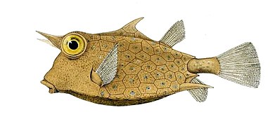
- The dorsal fin spines of are fused and modified placoid scales.
- caudal peduncle.[56]
- Some anchovies, and halfbeakshave deciduous scales, which are easily shed and aid in escaping predators.
- Male Percina darters have a row of enlarged caducous scales between the pelvic fins and the anus.
- Porcupine fishes have scales modified into large external spines.
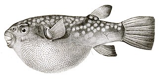
- By contrast, pufferfish have thinner, more hidden spines than porcupine fish, which become visible only when the fish puffs up. Unlike the porcupine fish, these spines are not modified scales, but develop under the control of the same network of genes that produce feathers and hairs in other vertebrates.[57][58]
-
 Porcupine fish have scales modified into spines.
 -
 Pufferfishspines are not modified scales but are developed by an independent gene network.


Fish without scales
-
 Mandarinfish lack scales and protect themselves with a layer of smelly and bitter slime.

Fish without scales usually evolve alternatives to the protection scales can provide, such as tough leathery skin or bony plates.
- Jawless fish (lampreys and hagfishes) have smooth skin without scales and without dermal bone.[59] Lampreys get some protection from a tough leathery skin. Hagfish exude copious quantities of slime or mucus if they are threatened.[60] They can tie themselves in an overhand knot, scraping off the slime as they go and freeing themselves from a predator.[61]
- Most eels are scaleless, though some species are covered with tiny smooth cycloid scales.
- Most catfish lack scales, though several families have body armour in the form of dermal plates or some sort of scute.[62]
- aposematic.[63]
- Anglerfish have loose, thin skin often covered with fine forked dermal prickles or tubercles, but they do not have regular scales. They rely on camouflage to avoid the attention of predators, while their loose skin makes it difficult for predators to grab them.
Many groups of bony fishes, including
- Seahorses lack scales but have thin skin stretched over a bony plate armour arranged in rings through the length of their bodies.
- In boxfish, the plates fuse together to form a rigid shell or exoskeleton enclosing the entire body. These bony plates are not modified scales but skin that has been ossified. Because of this heavy armour boxfish are limited to slow movements, but few other fish are able to eat the adults.

| |
Eels seem scaleless, but some species are covered with tiny smooth cycloid scales.
| |
-
 Boxfishhave plates of ossified skin fused together to form a rigid shell.
 -
 Seahorses have thin skin stretched over bony plates arranged in rings.


Some fish, such as hoki and swordfish, are born with scales but shed them as they grow.
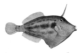
Filefish have rough non-overlapping scales with small spikes, which is why they are called filefish. Some filefish appear scaleless because their scales are so small.
Prominent scaling appears on tuna only along the lateral line and in the corselet, a protective band of thickened and enlarged scales in the shoulder region. Over most of their body tuna have scales so small that to casual inspection they seems scaleless.[64]
-
 Some filefish appear scaleless because their scales are so small.
 -
 To casual examination tuna seem largely free of scales, but they are not.


Leviticus
A passage in
According to the chok or divine decrees of the
Lepidophagy

Fish scales can be nutritious, containing a dermal portion and a layer of protein-rich mucus apart from the layers of
See also
- Age determination in fish
- Animal coloration
- Animal reflectors
- Photonic crystals
- Reptile scale
- Scale (zoology)
- Scale armour
- Snake scales
- Urokotori – Japanese fish scaler
References
- ^ Scale Etymonline. Retrieved 28 April 2019.
- PMID 23660349.
- S2CID 18868124.
- .
- S2CID 85993241.
- ^ ISBN 978-0-19-854047-2.
- ^ a b Turner, S. (1991). "Monophyly and interrelationships of the Thelodonti". In M. M. Chang; Y. H. Liu; G. R. Zhang (eds.). Early Vertebrates and Related Problems of Evolutionary Biology. Science Press, Beijing. pp. 87–119.
- .
- S2CID 128939911.
- PMID 28241029.
- ^ MICHAEL ALLABY "cosmoid scale ." A Dictionary of Zoology . . Encyclopedia.com. 29 Oct. 2019 <https://www.encyclopedia.com>
- ^ Zylberberg, L., Meunier, F.J., Laurin, M. (2010). A microanatomical and histological study of the postcranial dermal skeleton in the Devonian sarcopterygian Eusthenopteron foordi, Acta Palaeontologica Polonica 55: 459–470.
- PMID 30761080.
- S2CID 21829206.
- ^ PMID 9294653.
- PMID 19422423.
- ^ .
- ^ PMID 18660814.
- ^ PMID 27974575.
- ^ "Missouri Alligator Gar Management and Restoration Plan" (PDF). Missouri Department of Conservation Fisheries Division. January 22, 2013. Archived from the original (PDF) on May 6, 2016. Retrieved April 12, 2019.
- ^ Lagler, K. F., J. E. Bardach, and R. R. Miller (1962) Ichthyology. New York: John Wiley & Sons.
- ISBN 978-1-118-92421-1.
- ^ McGrouther, Mark (2 December 2019). "Cycloid and Ctenoid Scales". The Australian Museum. Retrieved 29 December 2021.
- ^ Pouyaud, L.; Sudarto, Guy G. Teugels (2003). "The different colour varieties of the Asian arowana Scleropages formosus (Osteoglossidae) are distinct species: morphologic and genetic evidences". Cybium. 27 (4): 287–305.
- ^ Ismail, M. (1989). Systematics, Zoogeography, and Conservation of the Freshwater Fishes of Peninsular Malaysia (Doctoral Dissertation ed.). Colorado State University.
- ^ E.J. Brill (1953). The Fishes of the Indo-Australian Archipelago. E.J. Brill. pp. 306–307.
- ^ a b c d Kawasaki, Kenta C., "A Genetic Analysis of Cichlid Scale Morphology" (2016). Masters Theses May 2014 - current. 425. http://scholarworks.umass.edu/masters_theses_2/425
- ^ Helfman, Gene (2009). The Diversity of Fishes Biology, Evolution, and Ecology. Wiley-Blackwell.
- ^ ISBN 9780198549567.
- ^ "There Are Probably Fish Scales In Your Lipstick". HuffPost India. 23 April 2015. Retrieved 6 May 2019.
- ^ Martin, R. Aidan. "Skin of the Teeth". Retrieved 28 August 2007.
- PMID 15667174.
- S2CID 18118663.
- ^ PMID 31137624.
- PMID 24943377.
- ^ Martin, R. Aidan. "The Importance of Being Cartilaginous". ReefQuest Centre for Shark Research. Retrieved 29 August 2009.
- ^ S2CID 122574419.
- ^ S2CID 23881820.
- ^ S2CID 137143652.
- ^ "Experimental investigations on drag-reduction characteristics of bionic surface with water-trapping microstructures of fish scales" (PDF).
- PMID 26064576.
- ^ .
- S2CID 139103148.
- PMID 25338940.
- ISSN 1369-7021.
- ^ PMID 22995249.
- ^ "Sharklet Discovery | Sharklet Technologies, Inc". www.sharklet.com. Retrieved 26 September 2018.
- PMID 22323201.
- ^ a b Dean, Brian & Bhushan, Bharat. (2010). Shark-Skin Surfaces for Fluid-Drag Reduction in Turbulent Flow: A Review. Philosophical transactions. Series A, Mathematical, physical, and engineering sciences. 368. 4775-806. 10.1098/rsta.2010.0201.
- ^ PMID 29436512.
- ^ S2CID 19556201.
- ^ PMID 15272388.
- ^ S2CID 13353402.
- ^ PMID 15272389.
- ^ PMID 10090147.
- PMID 23542000.
- ^ How the pufferfish got its wacky spines Phys.org, 25 July 2019.
- PMID 31353167.
- ISBN 9780080549521
- ^ Rothschild, Anna (1 April 2013). "Hagfish slime: The clothing of the future?". BBC News. Retrieved 2 April 2013.
- ^ Yong, Ed (23 January 2019). "No One Is Prepared for Hagfish Slime". The Atlantic. Retrieved 26 January 2019.
- JSTOR 1447528.
- .
- ^ Do tunas have scales? Northeast Fisheries Science Center, NOAA Fisheries. Accessed 4 August 2019.
- ^ Leviticus 11:9–10
- ^ a b c Aryeh Citron, "All About Kosher Fish"
- ^ Verifying Kosher Fish OU Kosher Certification. Retrieved 9 August 2019.
- ^ PMID 22970282.
- ^ Froese, R. and D. Pauly. Editors. "Glossary: Lepidophagy". FishBase. Retrieved 12 April 2007.
{{cite web}}:|author=has generic name (help) - ^ S2CID 15566769.
- S2CID 23695342.
- S2CID 33113282.
- PMID 20102595.
Further reading
- Helfman, G.S., B.B. Collette and D.E. Facey (1997). The Diversity of Fishes. Blackwell Science. pp. 33–36. ISBN 978-0-86542-256-8.)
{{cite book}}: CS1 maint: multiple names: authors list (link - Schultze, H.P. (2016). "Scales, enamel, cosmine, ganoine, and early osteichthyans". Comptes Rendus Palevol. 15 (1–2): 83–102. .