
Mallory body

In
intermediate filaments
within the liver cells.
Associated conditions
Mallory bodies are classically found in the
alcohol-induced liver disease and were once thought to be specific for that.[2]
They are most common in
alcoholic cirrhosis (prevalence of 51%).[3]
They are a recognized feature of
morbid obesity (8%), among other conditions.[3] However, it has also been reported in certain other unrelated conditions.[4]
Appearance
Mallory bodies are highly
proteins that have been ubiquitinated, or bound by other proteins such as heat shock proteins, or p62/Sequestosome 1.[5]
Eponym
It is named for the American pathologist Frank Burr Mallory, who first described the structures in 1911.[3] A renaming as Mallory–Denk bodies was proposed in 2007 to honor the contribution of Austrian pathologist Helmut Denk for the molecular analysis of the pathogenesis of MDBs.[6]
See also
- Ballooning degeneration – another histopathologic finding of steatohepatitis.
Additional images
-
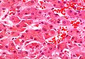
 Micrograph showing a Mallory body. Original magnification 400X. H&E stain.
 -
 Micrograph showing a Mallory body. Original magnification 200X. H&E stain.
 -
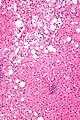
 Liver micrograph showing abundant Mallory bodies, as seen in alcohol use disorder.
 -
 Mallory bodies inTrichrome stain.


