
Median sacral artery
| Median sacral artery | |
|---|---|
 The abdominal aorta and its branches. (Middle sacral visible at center bottom.) | |
 The arteries of the pelvis. (Middle sacral labeled at upper right.) | |
| Details | |
| Source | abdominal aorta |
| Vein | Median sacral vein |
| Supplies | coccyx, lumbar vertebrae, sacrum |
| Identifiers | |
| Latin | arteria sacralis mediana |
| TA98 | A12.2.12.008 |
| TA2 | 4298 |
| FMA | 14757 |
| Anatomical terminology | |
The median sacral artery (or middle sacral artery) is a small artery that arises posterior to the abdominal aorta and superior to its bifurcation.
Structure
The median sacral artery arises from the
glomus coccygeum
(coccygeal gland).
Minute branches pass from it, to the posterior surface of the rectum.
On the last lumbar vertebra it
anterior sacral foramina
.
It is crossed by the left
venae comitantes; these unite to form a single vessel that opens into the left common iliac vein
.
Development
The median sacral artery is morphologically the direct continuation of the abdominal aorta.[2] It is vestigial in humans, but large in animals with tails, such as the crocodile.
See also
Additional images
-
 The iliac veins.
 -
 Scheme of the anastomosis of the veins of the rectum.
 -
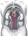 The relations of the viscera and large vessels of the abdomen.
 -
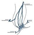
 Median sacral artery
 -
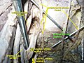
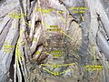 Pelvic contents: male. Superior view. Deep dissection.
 -
 Median Sacral Artery


References
![]() This article incorporates text in the public domain from page 613 of the 20th edition of Gray's Anatomy (1918)
This article incorporates text in the public domain from page 613 of the 20th edition of Gray's Anatomy (1918)
External links
- Anatomy photo:40:11-0200 at the SUNY Downstate Medical Center - "Posterior Abdominal Wall: Branches of the Abdominal Aorta"
- pelvis at The Anatomy Lesson by Wesley Norman (Georgetown University) (pelvicarteries)