
Tympanic cavity
| Tympanic cavity | |
|---|---|
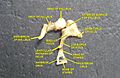
 The cochlea and vestibule, viewed from above. (Tympanic cavity labeled at upper right.) | |

Tensor tympani Stapedius Labyrinth Tympanic membrane (Ear drum) Tympanic cavity Bones and muscles in the tympanic cavity in the middle ear | |
| Details | |
pharyngeal pouch | |
| Part of | Middle ear |
| Artery | stylomastoid artery |
| Identifiers | |
| Latin | cavitas tympani |
| TA98 | A02.1.06.051 |
| TA2 | 687 |
| FMA | 56461 |
| Anatomical terminology] | |
The tympanic cavity is a small cavity surrounding the bones of the middle ear. Within it sit the ossicles, three small bones that transmit vibrations used in the detection of sound.
Structure
On its lateral surface, it abuts the
tympanic membrane
(eardrum).
Walls
The tympanic cavity is bounded by:
- Facing the prominence of the facial canal.
- Facing the internal maxillary artery. The iter chordæ anterius (canal of Huguier) is placed at the medial end of the petrotympanic fissure; through it the chorda tympani nerve leaves the tympanic cavity.
- The roof of the cavity (also called the tegmental wall, tegmental roof or tegmentum tympani) is formed by a thin plate of bone, the tegmen tympani, which separates the tympanic antrum, and forward to cover in the semicanal for the tensor tympani muscle. Its lateral edge corresponds with the remains of the petrosquamous suture.[1] The Atticus is the part of the tegmentum tympani where the stapes and incusare attached.
- The floor of the cavity (also called the jugular wall) is narrow, and consists of a thin plate of bone (fundus tympani) which separates the tympanic cavity from the jugular fossa. It presents, near the labyrinthic wall, a small aperture for the passage of the tympanic branch of the glossopharyngeal nerve.
- The posterior wall (or mastoid wall) is wider above than below, and presents for examination the entrance to the tympanic antrum, the pyramidal eminence, and the fossa incudis.
- The anterior wall (or carotid wall) is wider above than below; it corresponds with the petrous portion of the temporal bone.
Development
It is formed from the
tubotympanic recess, an expansion of the first pharyngeal pouch
.
Clinical significance
If damaged, the
tympanic membrane can be repaired in a procedure called tympanoplasty
.
Should fluid accumulate within the middle ear as the result of infection or for some other reason, it can be drained by puncturing the tympanic membrane with a large bore needle (tympanocentesis).
Additional images
-
 External and middle ear, opened from the front. Right side.
 -
 Horizontal section through left ear; upper half of section.
 -
 Tympanic cavity. Facial canal. Internal carotid artery.
 -
 Auditory ossicles. Tympanic cavity. Deep dissection.


References
![]() This article incorporates text in the public domain from page 1037 of the 20th edition of Gray's Anatomy (1918)
This article incorporates text in the public domain from page 1037 of the 20th edition of Gray's Anatomy (1918)
- ^ Public domain edition of Gray's Anatomy
External links
Wikimedia Commons has media related to Tympanic cavity.