
Ovine rinderpest
| Morbillivirus caprinae | |
|---|---|
| Virus classification | |
| (unranked): | Virus |
| Realm: | Riboviria |
| Kingdom: | Orthornavirae |
| Phylum: | Negarnaviricota
|
| Class: | Monjiviricetes |
| Order: | Mononegavirales |
| Family: | Paramyxoviridae |
| Genus: | Morbillivirus |
| Species: | Morbillivirus caprinae
|
| Synonyms[1] | |
| |
Ovine rinderpest, also commonly known as peste des petits ruminants (PPR), is a contagious disease primarily affecting goats and sheep; however, camels and wild small ruminants can also be affected.[2] PPR is currently present in North, Central, West and East Africa, the Middle East, and South Asia.[3] It is caused by Morbillivirus caprinae in the genus Morbillivirus, and is closely related to, among others, Morbillivirus pecoris, Morbillivirus hominis (Measles virus), and Morbillivirus canis (also known as canine distemper virus). The disease is highly contagious, and can have an 80–100% mortality rate in acute cases in an epizootic setting. The virus does not infect humans.
The disease was first described in 1942 in
In 2017, the disease was reported to be affecting saiga antelope in Mongolia, causing near-catastrophic herd depletion for the endangered species.[5]
In 2018, it was stated that the disease was reported to be present in Bulgaria close to the border with Turkey.[6] In a flock of 540 sheep and goats, two animals tested positive and one died, with disease confirmed by only one laboratory without any further tests.[7] Nevertheless, over 4000 sheep and goats were killed.[8]
Synonyms
PPR is also known as goat plague, kata, syndrome of stomatitis-pneumoenteritis, and ovine rinderpest.[9]
Official agencies such as the
Signs and symptoms
Symptoms are similar to those of rinderpest in cattle and involves oral necrosis, mucopurulent nasal and ocular discharges, cough, pneumonia, and diarrhea,[10] though they vary according to the previous immune status of the sheep, the geographic location, the time of year, or if the infection is new or chronic. They also vary according to the breed of sheep. However, fever in addition to either diarrhea or signs of oral discomfort is sufficient to suspect the diagnosis.[10] Incubation period is 3–5 days.[11]
Hyperacute cases
Hyperacute cases are found dead without previous symptoms. They die with a serous, foamy, or haemorrhagic discharge coming out of the nose.
Acute cases at onset
In acute cases, animals are recumbent, sometimes in self-auscultation position.
Evolution of acute cases
Nasal discharge becomes
-
 Self-auscultation in an acute case
 -
 Hind legs stained with sticky diarrhoea
 -
 Arched back (painful defecation)
 -
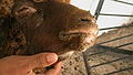
 inflammation and erosion of the mouth
 -
 Periodontitis
 -
 Mucopurulent nasal exudate
 -
 Orf-like scabs on lips in a recovering case, Day 8


Cause
Peste des petits ruminants is caused by a
Epidemiology
Origin and spread
This virus appears to have evolved at the start of the 20th century in Nigeria.[16] The extant genotypes subsequently appeared in West Africa (lineages I and II), East Africa and Arabia (lineage III), and Pakistan–India (lineage IV).[10]
The first description of the disease was published in 1942 and relates to an outbreak in
The outbreak in Burkina Faso in 1999 was caused by the lineage I group. Genotype III has caused outbreaks in Ethiopia (1996) and also in Arabia, southern India, and Tamil Nadu (1992). This lineage was found in Yemen in 2001. Genotype IV has been isolated in Kuwait in 1999.
Geographical repartition
As of 2017, the disease is present in West Africa, part of Central Africa (Gabon, Central African Republic), East Africa (north of the Equator), the Middle East and the Indian subcontinent including Nepal and Myanmar. The disease is endemic in the Indian subcontinent and is a major threat to fast-growing goat husbandry in India, causing an annual loss of around 1800 million Indian rupees.
In North Africa, only Egypt was once hit, but since summer 2008, Morocco is suffering a generalized outbreak with 133 known cases in 129 provinces, mostly affecting sheep.[18] The outbreak has precipitated the vaccination of a large number of the 17 million sheep and five million goats in the country.[19]
Dissemination
The disease is transmitted by infected
In an affected flock, even in pest-free regions, the disease does not progress very rapidly, in spite of the close contact between animals. New clinical cases may be observed daily for a 1-month period.[20]
Post mortem lesions
The lesions are situated in the digestive tract. Quick post mortem examination will lead to the discovery of many haemorrhagic patches on the
Erosions and inflammation are widespread on buccal mucosa. The same lesions are also present in
Microscopic lesions
Microscopic study of both natural and experimental cases revealed
]In summary Epithelial cells, alveolar macrophages, lungs, and hepatocytes all showed histopathological alterations, primarily infiltrations of inflammatory cells, syncytia, and presence of intranuclear and/or intracytoplasmic eosinophillic inclusions.[23]
Diagnosis
History and clinical signs enable a presumptive diagnosis to be made in endemic regions. The virus can be detected in acute cases from various swabs and blood samples, using PCR and ELISA. Antibodies can also be detected by ELISA.[14]
Treatment and control
Antibiotics such as chloramphenicol, penicillin, and streptomycin can be used and supportive treatment may be helpful.[14] Additionally, a vaccine has been developed that may decrease death in the flock.[14] Movement restrictions and slaughter of affected flocks may be required in an attempt to eradicate the disease.[14] A global eradication programme has been developed by the Food and Agriculture Organization of the United Nations and the World Organisation for Animal Health.[24] More information can be found on FAO's website on the implementation of this global plan.[25] It is considered feasible to eradicate ovine rinderpest in 15 years, starting in the year 2016.[10]
References
- ^ "ICTV Taxonomy history: Small ruminant morbillivirus". International Committee on Taxonomy of Viruses (ICTV). Retrieved 15 January 2019.
- PMID 23305511.
- PMID 20844089.
- ^ "Peste des Petits Ruminants". Food and Agriculture Organization of The United Nations.
- ^ "PESTE DES PETITS RUMINANTS – MONGOLIA (03): (HOVD) SAIGA ANTELOPE". ProMED-mail. 9 March 2017. Retrieved 9 March 2017.
- ^ "Bulgaria reports another case of ovine rinderpest". Reuters. 19 July 2018. Retrieved 2 August 2018.
- ^ Prevalence of Antimicrobial Resistance in Ruminants in Bulgaria: A Pilot Study
- ^ Protests amid outrage over killing of sheep and goats over rinderpest outbreak in Bulgaria
- PMID 26443889.
- ^ S2CID 255107410.
- ^ "Rinderpest | animal disease". Encyclopedia Britannica. Retrieved 2019-12-07.
- ^ J. Berrada, Observations des premiers cas confirmés de peste des petits ruminants au Maroc, oral presentation, El Jadida, 31-07-2008.
- ^ Handbook of Animal Diseases in the Tropics, op cit.
- ^ a b c d e Peste des Petits Ruminants Archived 2013-02-22 at archive.today reviewed and published by WikiVet, accessed 10 October 2011.
- PMID 21762576.
- PMID 25418782.
- PMID 12135634.
- FAO. September 9, 2008. Retrieved 2008-09-10.
- ^ "Morocco to vaccinate all livestock after virus outbreak". AFP. September 9, 2008. Archived from the original on May 20, 2011. Retrieved 2008-09-10.
- ^ L. Mahin (2008) Observations sur un foyer de Peste des petits ruminants, unpublished data.
- ^ Tligui, Observations nécropsiques sur les premiers cas confirmés de peste des petits ruminants au Maroc, oral presentation, El Jadida, 31-07-2008.
- ^
Seki, Fumio; Takeda, Makoto (2022). "Novel and classical morbilliviruses: Current knowledge of three divergent morbillivirus groups". Microbiology and Immunology. 66 (12): 552–563. S2CID 252497033. This review cites this research. Sahoo, Monalisa; M, Dinesh; Thakor, Jigarji Chaturji; Baloni, Suraj; Saxena, Sonal; Shrivastava, Sameer; Dhama, Kuldeep; Singh, Karampal; Singh, Rajendra (2020-03-01). "Neuropathology mediated through caspase dependent extrinsic pathway in goat kids naturally infected with PPRV".S2CID 209482267.
- ^
Maes, Piet; et al. (2018). "Taxonomy of the family Arenaviridae and the order Bunyavirales: Update 2018". Archives of Virology. 163 (8): 2295–2310. S2CID 254054051. This review cites this research. Blasdell, Kim R.; Duong, Veasna; Eloit, Marc; Chretien, Fabrice; Ly, Sowath; Hul, Vibol; Deubel, Vincent; Morand, Serge; Buchy, Philippe (2016). "Evidence of human infection by a new mammarenavirus endemic to Southeastern Asia". eLife. 5.PMID 27278118.
- ^ FAO and WHO. 2016. Peste des petits ruminants GLOBAL ERADICATION PROGRAM. Rome.
- ^ FAO. Peste des Petits Ruminants. http://www.fao.org/ppr/en/ accessed Nov. 2016
External links
- PPR Global Eradication Program by FAO
- Peste-des-Petits-Ruminants at the U.S. National Library of Medicine Medical Subject Headings (MeSH) (disease)
- Peste-des-petits-ruminants+virus at the U.S. National Library of Medicine Medical Subject Headings (MeSH) (pathogen)
- Current status of Ovine rinderpest (Peste des petits ruminant PPR) worldwide at OIE. WAHID Interface – OIE World Animal Health Information Database
- Disease card
- WikiVet summary of disease and links to key references on CABI
- The disease in the Merck veterinary manual Archived 2016-03-03 at the Wayback Machine
- Field manual for recognition, at fao.org
- Overview at vet.uga.edu
- Virus Phylogenetic Tree at bbsrc.ac.uk