
Superior vena cava
| Superior vena cava | |
|---|---|
Right atrium | |
| Identifiers | |
| Latin | vena cava superior, vena maxima |
| MeSH | D014683 |
| TA98 | A12.3.03.001 |
| TA2 | 4745 |
| FMA | 4720 |
| Anatomical terminology] | |
The superior vena cava (SVC) is the superior of the two venae cavae, the great venous trunks that return deoxygenated blood from the systemic circulation to the right atrium of the heart. It is a large-diameter (24 mm) short length vein that receives venous return from the upper half of the body, above the diaphragm. Venous return from the lower half, below the diaphragm, flows through the inferior vena cava. The SVC is located in the anterior right superior mediastinum.[1] It is the typical site of central venous access via a central venous catheter or a peripherally inserted central catheter. Mentions of "the cava" without further specification usually refer to the SVC.[citation needed]
Structure
The superior vena cava is formed by the left and right
The superior vena cava is made up of three layers, starting with the innermost endothelial
Anatomical variation
The most common anatomical variation is a persistent left superior vena cava. In persons with a persistent left superior vena cava, the right superior vena cava may be normal, small or absent, with or without an anterior communicating vein. This variation is present in less than 0.5% of the general population, but in up to 10% in patients with congenital heart disease.[5]
Clinical significance
In
No valve divides the superior vena cava from the right atrium. As a result, the (right) atrial and (right) ventricular contractions are conducted up into the internal jugular vein and, through the sternocleidomastoid muscle, can be seen as the jugular venous pressure.
Additional images
-
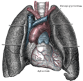 The thorax, viewed from the front, showing the superior vena cava between the heart and lungs.
 -
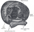 Heart seen from above, with the valve-less entry of the superior vena cava visible on the right.
 -
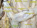 Superior vena cava in a cadaveric specimen.
 -
 Cross-section of the thorax showing the formation of the superior vena cava.


See also
References
- ^ "General Practice Notebook". www.gpnotebook.co.uk. Retrieved April 6, 2018.
- PMID 31424839, retrieved November 24, 2021
- PMID 8562136.
- ^ Zhang, Shu-Xin (1999). An Atlas of Histology. New York: Springer. pp. 131–133.
- PMID 26452112.
- ISBN 978-0-7020-3085-7.)
{{cite book}}:|first=has generic name (help)CS1 maint: multiple names: authors list (link
| National | |
|---|---|
| Other | |