
Tricuspid valve
| Tricuspid valve | |
|---|---|
.svg) Anterior (frontal) view of the opened heart. White arrows indicate normal blood flow. (Tricuspid valve labeled at bottom left.) | |
 Heart in motion: the anterior walls of the ventricles are removed. The action of the tricuspid valve, located in the right ventricle, is seen on the left portion of this illustration. The three leaflets with their attached chordae tendineae and papillary muscles can be seen. | |
| Details | |
| Identifiers | |
| Latin | valvula tricuspidalis, valva atrioventricularis dextra |
| MeSH | D014261 |
| TA98 | A12.1.02.003 |
| TA2 | 3982 |
| FMA | 7234 |
| Anatomical terminology | |
The tricuspid valve, or right atrioventricular valve, is on the right dorsal side of the mammalian
Structure
The tricuspid valve usually has three
Function
The tricuspid valve functions as a one-way valve that closes during
Clinical significance
Infected valves can result in endocarditis in intravenous drug users.[5][6] Patients who inject narcotics or other drugs intravenously may introduce infection, which can travel to the right side of the heart, most often caused by the bacteria S. aureus.[7] In patients without a history of intravenous exposure, endocarditis is more frequently left-sided.[7]
The tricuspid valve can be affected by
Certain carcinoid syndromes can affect the tricuspid valve by producing fibrosis due to serotonin production by those tumors.
The first endovascular tricuspid valve implant was performed by surgeons at the Cleveland Clinic.[9]
Tricuspid regurgitation
Additional images
-
 Tricuspid valve. Deep dissection.
 -
 Tricuspid valve marked in yellow.
 -
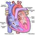 Diagram of tricuspid insufficiency/regurgitation. Marked in black arrow.


See also
References
- ^ "Anatomy of the Tricuspid Valve". e-echocardiography.com. Retrieved 2018-03-30.
- ^ Richard Van Pragh: Cardiac anatomy in A. C. Chang et al.: Pediatric Cardiac Intensive Care, Philadelphia 1998.
- PMID 10430758.
- ^ "Enlarged heart - Symptoms and causes". Mayo Clinic. Retrieved 2018-03-30.
- PMID 11019526.
- S2CID 32603989.
- ^ ISBN 978-1-4160-2973-1.
- Mount Sinai Hospital, New York
- ^ University Circle Inc. Archived 2008-06-17 at the Wayback Machine
- ^ PMID 26358570.
- ^ Prihadi', 'Edgard A. "Tricuspid valve regurgitation: no longer the "forgotten valve"". www.escardio.org. Retrieved 2021-11-27.
External links
- Anatomy figure: 20:07-04 at Human Anatomy Online, SUNY Downstate Medical Center
- Photo of model: circulation/tricuspidvalve04 at Waynesburg College
- Cardiac Valve Animations - Perioperative Interactive Education Group