
Sinoatrial node
| Sinoatrial node | |
|---|---|
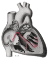
Electrical conduction system of the heart | |
| Artery | Sinoatrial nodal artery |
| Identifiers | |
| Latin | nodus sinuatrialis |
| Acronym(s) | SA node |
| MeSH | D012849 |
| TA98 | A12.1.06.003 |
| TA2 | 3953 |
| FMA | 9477 |
| Anatomical terminology] | |
The sinoatrial node (also known as the sinuatrial node, SA node or sinus node) is an
These cells can produce an
Structure
The sinoatrial node is an oval-shaped structure that is approximately 15 mm long, 3 mm wide, and 1 mm thick, located directly below and to the side of the superior vena cava.[1] The size can vary but is usually between 10-30 mm long, 5–7 mm wide, and 1–2 mm deep.[3][4]
Location
The SA node is located in the wall (
Microanatomy

The cells of the SA node are spread out within a mesh of
Action potentials pass from one cardiac cell to the next through pores known as gap junctions. These gap junctions are made of proteins called connexins. There are fewer gap junctions within the SA node and they are smaller in size. This is again important in insulating the SA node from the surrounding atrial cells.[2][8]
Blood supply
The sinoatrial node receives its blood supply from the sinoatrial nodal artery. This blood supply, however, can differ hugely between individuals. For example, in most humans, this is a single artery, although in some cases there have been either 2 or 3 sinoatrial node arteries supplying the SA node. Also, the SA node artery mainly originates as a branch of the right coronary artery; however in some individuals it has arisen from the circumflex artery, which is a branch of the left coronary artery. Finally, the SA node artery commonly passes behind the superior vena cava, before reaching the SA node; however in some instances it passes in front. Despite these many differences, there doesn't appear to be any advantage to how many sinoatrial nodal arteries an individual has, or where they originate.[9]
Venous drainage
There are no large
Function
Pacemaking
The main role of a sinoatrial node cell is to initiate action potentials of the heart that can pass through
Outlined below are the 3 phases of a sinoatrial node action potential. In the cardiac action potential, there are 5 phases (labelled 0-4), however pacemaker action potentials do not have an obvious phase 1 or 2.
Phase 4

This phase is also known as the
Phase 0
This is the depolarization phase. When the membrane potential reaches the threshold potential (around -20 to -50 mV), the cell begins to rapidly depolarise (become more positive).[16] This is mainly due to the flow of Ca2+ through L-type calcium channels, which are now fully open. During this stage, T-type calcium channels and HCN channels deactivate.
Phase 3
This phase is the repolarization phase. This occurs due to the inactivation of L-type calcium channels (preventing the movement of Ca2+ into the cell) and the activation of potassium channels, which allows the flow of K+ out of the cell, making the membrane potential more negative.[17]
Nerve supply
Heart rate depends on the rate at which the sinoatrial node produces action potentials. At rest, heart rate is between 60 and 100 beats per minute. This is a result of the activity of two sets of nerves, one acting to slow down action potential production (these are parasympathetic nerves) and the other acting to speed up action potential production (sympathetic nerves).[18]
Modulation of heart rate by ANS is carried by two types of channel: Kir and HCN (members of the CNG gated channels).
The sympathetic nerves begin in the
The
The first cell to produce the action potential in the SA node isn't always the same; this is known as pacemaker shift. In certain species of animals—for example, in dogs—a superior shift (i.e., the cell that produces the fastest action potential in the SA node is higher than previously) usually produces an increased heart rate whereas an inferior shift (i.e. the cell producing the fastest action potential within the SA node is further down than previously) produces a decreased heart rate.[2]
Clinical significance
Sinus node dysfunction also known as sick sinus syndrome is a group of irregular heartbeat conditions caused by faulty electrical signals of the heart. When the heart's sinoatrial node is defective, the heart's rhythms become abnormal—typically too slow or exhibiting pauses in its function or a combination, and very rarely faster than normal.[21]
Blockage of the arterial blood supply to the SA node (most commonly due to a myocardial infarction or progressive coronary artery disease) can therefore cause ischemia and cell death in the SA node. This can disrupt the electrical pacemaker function of the SA node, and can result in sinus node dysfunction.
If the SA node does not function or the impulse generated in the SA node is blocked before it travels down the electrical conduction system, a group of cells further down the heart will become its pacemaker.[22]
History
The sinoatrial node was first discovered by a young medical student, Martin Flack, in the heart of a mole, whilst his mentor, Sir Arthur Keith, was on a bicycle ride with his wife. They made the discovery in a makeshift laboratory set up in a farmhouse in Kent, England, called Mann's Place. Their discovery was published in 1907.[23][24]
Additional images
-
 Heart; conduction system (SA node labeled 1)
 -
 Schematic representation of the atrioventricular bundle


See also
- Cardiac pacemaker
- Cardiology
- Heart block
- Sinus bradycardia
- Sinus tachycardia
- Cardiothoracic Surgery
References
- ^ ISBN 9781416045748.)
{{cite book}}: CS1 maint: location missing publisher (link - ^ S2CID 22207608.
- PMID 26743207.
- S2CID 45464625.
- ^ Elsevier, Dorland's Illustrated Medical Dictionary, Elsevier.
- PMID 19289639.
- PMID 8930845.
- ^ PMID 10974216.
- PMID 26849441.
- PMID 426954.
- PMID 350631.
- PMID 330018.
- PMID 8380502.
- PMID 20167941.
- PMID 21319337.
- ^ Verkerk, A., Borren, van, Peters, R., Broekhuis, E., Lam, K., Coronel, R., Bakker, de, Tan, H. and Wilders, R. (2007) 'Single cells isolated from human sinoatrial node: Action potentials and numerical reconstruction of pacemaker current', Conference proceedings : ... Annual International Conference of the IEEE Engineering in Medicine and Biology Society. IEEE Engineering in Medicine and Biology Society. Annual Conference., 2007, pp. 904–7.
- S2CID 476037.
- PMID 25914789.
- ^ Larsson, P.H. (2010) 'How is the heart rate regulated in the sinoatrial node? Another piece to the puzzle', 136(3).
- ^ Osterrieder W., Noma A., Trautwein W. (1980) On the kinetics of the potassium current activated by acetylcholine in the SA node of the rabbit heart. Pflügers Arch. 386:101–109.
- Mount Sinai Hospital, New York
- ^ Junctional Rhythm at eMedicine
- PMID 17890694.
- S2CID 17882001.
External links
- Anatomy figure: 20:06-01 at Human Anatomy Online, SUNY Downstate Medical Center - "The conduction system of the heart."
- Diagram at gru.net
- thoraxlesson4 at The Anatomy Lesson by Wesley Norman (Georgetown University) (thoraxheartinternalner)
- https://web.archive.org/web/20070929080346/http://www.healthyheart.nhs.uk/heart_works/heart03.shtml