
Eustachian tube
| Eustachian tube | |
|---|---|
 External and middle ear. Eustachian tube labelled as auditory tube. | |
 Middle ear, with auditory tube at bottom right | |
| Details | |
| Pronunciation | /juːˈsteɪʃən/ |
| Precursor | First pharyngeal pouch |
| Identifiers | |
| Latin | tuba auditiva, tuba auditivea, tuba auditoria |
| MeSH | D005064 |
| TA98 | A15.3.02.073 |
| TA2 | 6926 |
| FMA | 9705 |
| Anatomical terminology | |
The Eustachian tube (
In humans and other
Structure


The Eustachian tube extends from the anterior wall of the middle ear to the lateral wall of the
Bony part
The bony part (1⁄3) nearest to the middle ear is made of bone and is about 12 mm in length. It begins in the anterior wall of the tympanic cavity, below the septum canalis musculotubarius, and, gradually narrowing, ends at the angle of junction of the squamous and the petrous parts of the temporal bone, its extremity presenting a jagged margin which serves for the attachment of the cartilaginous part.[5] The vestibule of the Eustachian tube is known as the protympanum,[6] The protympanum is also known as the anterior part of the bony part of the tube.[7]
Cartilaginous part
The cartilaginous part of the Eustachian tube is about 24 mm in length and is formed of a triangular plate of
The upper edge of the
The position and relations of the pharyngeal opening are described with the
Muscles
There are four muscles associated with the function of the Eustachian tube:
- Levator veli palatini (innervated by the vagus nerve)
- Salpingopharyngeus(innervated by the vagus nerve)
- Tensor veli palatini(innervated by the mandibular nerve of CN V)
The tube is opened during swallowing by contraction of the tensor veli palatini and levator veli palatini, muscles of the soft palate.[1] The tensor veli palatini makes the largest contribution to active opening of the tube.[9][10]
New anatomical perspectives
Since 2015, two developments have enhanced our understanding of the anatomy of the eustachian tube: Valsalva computerized tomography and endoscopic ear surgery.[11]
- Given the greater access to the ear anatomy using endoscopic methods, it has been suggested that the bony part of the eustachian tube is really the anterior extension of the middle ear cavity, or the "Protympanum". The term "Eustachian Tube" should be limited to the fibrocartilaginous structure connecting the protympanum to the nasopharynx.[4]
- The Eustachian tube is a saclike irregular structure rather than a tubular structure.
- The ear side of the eustchian tube is by far the narrowest segment, called isthmus, and is probably the site of possible obstructive pathology causing chronic ear disease.[12]
Development
The Eustachian tube is derived from the dorsal part of the first
-
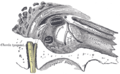
 Frontal section through left ear; upper half of section
 -
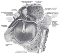
 View of the inner wall of the tympanum (enlarged)
 -
 The right membrana tympani with the hammer and the chorda tympani, viewed from within, from behind, and from above


Function
Pressure equalization
Under normal circumstances, the human Eustachian tube is closed, but it can open to let a small amount of air through to prevent damage by equalizing pressure between the middle ear and the atmosphere. Pressure differences cause temporary
Mucus drainage
The Eustachian tube also drains mucus from the middle ear. Upper respiratory tract infections or allergies can cause the Eustachian tube, or the membranes surrounding its opening to become swollen, trapping fluid, which serves as a growth medium for bacteria, causing
Clinical significance
Otitis media, or inflammation of the middle ear, commonly affects the Eustachian tube. Children under 7 are more susceptible to this condition, one theory being that this is because the Eustachian tube is shorter and at more of a horizontal angle than in the adult ear. Others argue that susceptibility in this age group is related to immunological factors and not Eustachian tube anatomy.[citation needed]
Barotitis, a form of barotrauma, may occur when there is a substantial difference in air or water pressure between the outer and the middle ear – for example, during a rapid ascent while scuba diving, or during sudden decompression of an aircraft at high altitude.
Some people are born with a dysfunctional Eustachian tube[16] that is much slimmer than usual. The cause may be genetic, but it has also been posited as a condition in which the patient did not fully recover from the effects of pressure on the middle ear during birth (retained birth compression).[17][unreliable medical source] It is suggested that Eustachian tube dysfunction can result in a large amount of mucus accumulating in the middle ear, often impairing hearing to a degree. This condition is known as otitis media with effusion.
A patulous Eustachian tube is a rare condition in which the Eustachian tube remains intermittently open, causing an echoing sound of the person's own heartbeat, breathing, and speech. This may be temporarily relieved by holding the head upside down.
Smoking can also cause damage to the
Recurring and chronic cases of sinus infection can result in Eustachian tube dysfunction caused by excessive mucus production which, in turn, causes obstruction to the openings of the Eustachian tubes.
Ventilation tubes
In severe cases of childhood middle ear infections and Eustachian tube blockage, ventilation can be provided by a surgical puncturing of the eardrum to permit air equalization, known as myringotomy. The eardrum would normally naturally heal and close the hole, so a tiny plastic rimmed grommet is inserted into the hole to hold it open. This is known as a tympanostomy tube. As a child grows, the tube is eventually naturally expelled by the body. Longer-lasting vent grommets with larger flanges have been researched, but these can lead to permanent perforation of the eardrum.
Dilation of the Eustachian tube
More recently, dilation of the eustachian tube using balloon catheter has gained attention as a method of treating eustachian tube obstruction. There are two methods of performing this procedure depending on the route of the catheter introduction and the area of the Eustachian tube to be dilated. Dennis Poe popularized the transnasal introduction and the dilation of the nose side of the eustachian tube.[19] Muaaz Tarabichi pioneered the transtympanic (ear) introduction of the balloon catheter and the dilatation of the proximal part (the ear side) of the cartilaginous eustachian tube.[20][21][22]
Other animals
In the
References
- ^ ISBN 978-1-4511-1945-9.
- ^ "Eustachian Tube Dysfunction or Blockage Symptoms & How to Clear". medicinenet.com. Archived from the original on 5 October 2017. Retrieved 6 May 2018.
- Who Named It?
- ^ PMID 27565395.
- University of Michigan Medical School
- ISBN 978-94-009-5002-3, retrieved 2024-01-16
- ^ Deng, Francis. "Protympanum | Radiology Reference Article | Radiopaedia.org". Radiopaedia.
- PMID 30085597, retrieved 2024-01-16
- ^ "The Eustachian (Auditory) Tube - Osseous - Cartilaginous -TeachMeAnatomy". teachmeanatomy.info. Retrieved 2023-11-18.
- ^ "Eustachian (auditory) tube". Kenhub. Retrieved 2023-11-18.
- S2CID 29964314.
- S2CID 7223491.
- ISBN 1-58890-173-4.
- PMID 7662078.
- ^ "Middle Ear, Eustachian Tube, Inflammation/Infection Treatment & Management". Medscape. Archived from the original on 2014-08-10. Retrieved 2014-08-06.
- ^ Eustachian Tube Function and Dysfunction Archived 2016-08-10 at the Wayback Machine at Baylor College of Medicine
- ^ "FAQs – Cranial Osteopathy". The Children's Clinic. Archived from the original on 2010-05-29. Retrieved 2008-12-23.
- S2CID 27862332.
- S2CID 4968887.
- S2CID 39239009.
- PMID 27565385.
- S2CID 52041093.
External links
| National | |
|---|---|
| Other | |