Filoviridae
| Filoviridae | |
|---|---|

| |
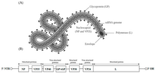
| Ebolavirus structure and genome | |

| |
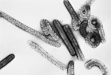
Electron micrograph of Marburg virus
| |
| Virus classification | |
| (unranked): | Virus |
| Realm: | Riboviria |
| Kingdom: | Orthornavirae |
| Phylum: | Negarnaviricota
|
| Class: | Monjiviricetes |
| Order: | Mononegavirales |
| Family: | Filoviridae |
| Genera | |
| |
Filoviridae (
All filoviruses are classified by the US as
Use of term
The
Note
According to the rules for taxon naming established by the International Committee on Taxonomy of Viruses (ICTV), the name Filoviridae is always to be capitalized, italicized, never abbreviated, and to be preceded by the word "family". The names of its members (filoviruses or filovirids) are to be written in lower case, are not italicized, and used without articles.[13][14]
Life cycle


The filovirus
Family inclusion criteria

A virus that fulfills the criteria for being a member of the order Mononegavirales is a member of the family Filoviridae if:[13][14]
- it causes viral hemorrhagic fever in certain primates
- it infects primates, pigs or bats in nature
- it needs to be adapted through serial passage to cause disease in rodents
- it exclusively replicates in the cytoplasm of a host cell
- it has a genome ≈19 kbp in length
- it has an RNA genome that constitutes ≈1.1% of the virion mass
- its genome has a molecular weight of ≈4.2×10⁶
- its genome contains one or more gene overlaps
- its genome contains seven genes in the order 3'-UTR-NP-VP35-VP40-GP-VP30-VP24-L-5'-UTR
- its VP24 gene is not homologous to genes of other mononegaviruses
- its genome contains transcriptioninitiation and termination signals not found in genomes of other mononegaviruses
- it forms nucleocapsids with a buoyant density in CsCl of ≈1.32 g/cm3
- it forms nucleocapsids with a central axial channel (≈10–15 nm in width) surrounded by a dark layer (≈20 nm in width) and an outer helical layer (≈50 nm in width) with a cross striation (periodicity of ≈5 nm)
- it expresses a class I fusion glycoprotein that is highly N- and O-glycosylated and acylated at its cytoplasmic tail
- it expresses a primary matrix protein that is not glycosylated
- it forms virions that bud from the plasma membrane
- it forms virions that are predominantly filamentous (U- and 6-shaped) and that are ≈80 nm in width, and several hundred nm and up to 14 μm in length
- it forms virions that have surface projections ≈7 nm in length spaced ≈10 nm apart from each other
- it forms virions with a molecular mass of ≈3.82×10⁸; an S20W of at least 1.40; and a buoyant density in potassium tartrate of ≈1.14 g/cm3
- it forms virions that are poorly neutralized in vivo
Family organization
| Genus name | Species name | Virus name (abbreviation) |
|---|---|---|
Cuevavirus
|
Lloviu cuevavirus
|
Lloviu virus (LLOV) |
Dianlovirus
|
Mengla dianlovirus | Měnglà virus (MLAV)
|
| Ebolavirus | Bombali ebolavirus | Bombali virus (BOMV) |
| Bundibugyo ebolavirus | Bundibugyo virus (BDBV; previously BEBOV)
| |
Reston ebolavirus
|
Reston virus (RESTV; previously REBOV) | |
| Sudan ebolavirus | Sudan virus (SUDV; previously SEBOV)
| |
| Taï Forest ebolavirus | Taï Forest virus (TAFV; previously CIEBOV)
| |
| Zaire ebolavirus | Ebola virus (EBOV; previously ZEBOV)
| |
| Marburgvirus | Marburg marburgvirus
|
Marburg virus (MARV) |
| Ravn virus (RAVV) | ||
| Striavirus | Xilang striavirus | Xīlǎng virus (XILV) |
| Thamnovirus | Huangjiao thamnovirus | Huángjiāo virus (HUJV) |
Phylogenetics
The mutation rates in these genomes have been estimated to be between 0.46 × 10⁻⁴ and 8.21 × 10⁻⁴ nucleotide substitutions/site/year.[15] The most recent common ancestor of sequenced filovirus variants was estimated to be 1971 (1960–1976) for Ebola virus, 1970 (1948–1987) for Reston virus, and 1969 (1956–1976) for Sudan virus, with the most recent common ancestor among the four species included in the analysis (Ebola virus, Tai Forest virus, Sudan virus, and Reston virus) estimated at 1000–2100 years.[16] The most recent common ancestor of the Marburg and Sudan species appears to have evolved 700 and 850 years before present respectively. Although mutational clocks placed the divergence time of extant filoviruses at ~10,000 years before the present, dating of orthologous endogenous elements (paleoviruses) in the genomes of hamsters and voles indicated that the extant genera of filovirids had a common ancestor at least as old as the Miocene (~16–23 million or so years ago).[17]
Filoviridae cladogram is the following:[18][19]
| Filoviridae |
| ||||||||||||||||||||||||||||||||||||||||||||||||||||||||||||||||||||||||||||||||||||||||||
Paleovirology
Filoviruses have a history that dates back several tens of million of years.
Vaccines
There are presently very limited vaccines for known filovirus.[24] An effective vaccine against EBOV, developed in Canada,[25] was approved for use in 2019 in the US and Europe.[26][27] Similarly, efforts to develop a vaccine against Marburg virus are under way.[28]
Mutation concerns and pandemic potential
There has been a pressing concern that a very slight genetic mutation to a filovirus such as
The
References
- ^ "Filoviridae". Merriam-Webster.com Dictionary. Retrieved July 28, 2018.
- PMID 31021739.
- ^ PMID 7118520.
- ^ US Animal and Plant Health Inspection Service (APHIS) and US Centers for Disease Control and Prevention (CDC). "National Select Agent Registry (NSAR)". Retrieved 2011-10-16.
- ^ US Department of Health and Human Services. "Biosafety in Microbiological and Biomedical Laboratories (BMBL) 5th Edition". Retrieved 2011-10-16.
- ^ US National Institutes of Health (NIH), US National Institute of Allergy and Infectious Diseases (NIAID). "Biodefense — NIAID Category A, B, and C Priority Pathogens". Archived from the original on 2011-10-22. Retrieved 2011-10-16.
- ^ US Centers for Disease Control and Prevention (CDC). "Bioterrorism Agents/Diseases". Archived from the original on July 22, 2014. Retrieved 2011-10-16.
- ^ The Australia Group. "List of Biological Agents for Export Control". Archived from the original on 2011-08-06. Retrieved 2011-10-16.
- ISBN 0-387-82286-0.
- ISBN 3-211-82594-0.
- ISBN 0-12-370200-3.
- ^ ISBN 0-12-370200-3.
- ^ PMID 21046175.
- ^ ISBN 978-0-12-384684-6.
- PMID 23255795.
- S2CID 9873900.
- PMID 25237605.
- PMID 34808081.)
{{cite journal}}: CS1 maint: multiple names: authors list (link - doi:10.1101/2023.08.07.552227.)
{{cite journal}}: CS1 maint: multiple names: authors list (link - PMID 20569424.
- PMID 20686665.
- PMID 21124940.
- PMID 22093762.
- PMID 9988154.
- PMID 29084758.
- ^ Research, Center for Biologics Evaluation and (2020-01-27). "ERVEBO". FDA.
- ^ CZARSKA-THORLEY, Dagmara (2019-10-16). "Ervebo". European Medicines Agency. Retrieved 2020-05-03.
- PMID 30567978.
- ^ Kelland, Kate (19 September 2014). "Scientists see risk of mutant airborne Ebola as remote". Reuters. Retrieved 10 October 2014.
- ^ "Feature Article: New Tech Makes Detecting Airborne Ebola Virus Possible". Department of Homeland Security. 20 April 2021. Retrieved 13 December 2021.
Further reading
- Klenk, Hans-Dieter (1999). Marburg and Ebola Viruses. Current Topics in Microbiology and Immunology. Vol. 235. Berlin, Germany: Springer-Verlag. ISBN 978-3-540-64729-4.
- Klenk, Hans-Dieter; Feldmann, Heinz (2004). Ebola and Marburg Viruses—Molecular and Cellular Biology. Wymondham, Norfolk, UK: Horizon Bioscience. ISBN 978-0-9545232-3-7.
- Kuhn, Jens H. (2008). Filoviruses—A Compendium of 40 Years of Epidemiological, Clinical, and Laboratory Studies. Archives of Virology Supplement. Vol. 20. Vienna, Austria: Springer. ISBN 978-3-211-20670-6.
- Ryabchikova, Elena I.; Price, Barbara B. (2004). Ebola and Marburg Viruses—A View of Infection Using Electron Microscopy. Columbus, Ohio, USA: Battelle Press. ISBN 978-1-57477-131-2.
External links
- ICTV Report: Filoviridae
- "Filoviridae". NCBI Taxonomy Browser. 11266.
- "FILOVIR". Scientific resources for research on filoviruses. Archived from the original on 2020-07-30. Retrieved 2014-08-08.
- Theoretical Evidence That The Ebola Virus Zaire Strain May Be Selenium-Dependent: A Factor In Pathogenesis And Viral Outbreaks? Taylor 1995
- Can Selenite Be An Ultimate Inhibitor Of Ebola And Other Viral Infections? Lipinski 2015 Archived 2020-11-19 at the Wayback Machine
- Many In West Africa May Be Immune To Ebola Virus New York Times
