Hemoglobin E
| Hemoglobin E disease | |
|---|---|
| Other names | Haemoglobin E |
 | |
| Crystal structure of Hemoglobin E mutant (Glu26Lys) PDB entry 1vyt. Alpha chain in pink, beta chain in red. The lysine mutation highlighted as white spheres. | |
| Specialty | Hematology |
Hemoglobin E (HbE) is an abnormal hemoglobin with a single point mutation in the β chain. At position 26 there is a change in the amino acid, from glutamic acid to lysine (E26K). Hemoglobin E is very common among people of Southeast Asian, Northeast Indian, Sri Lankan and Bangladeshi descent.[1][2]
The βE mutation affects β-gene expression creating an alternate splicing site in the mRNA at codons 25-27 of the β-globin gene. Through this mechanism, there is a mild deficiency in normal β mRNA and production of small amounts of anomalous β mRNA. The reduced synthesis of β chain may cause
Hemoglobin E disease (EE)

Hemoglobin E disease results when the offspring inherits the gene for HbE from both parents. At birth, babies
Hemoglobin E trait: heterozygotes for HbE (AE)
Sickle-Hemoglobin E Disease (SE)
Compound heterozygotes with sickle-hemoglobin E disease result when the gene of hemoglobin E is inherited from one parent and the gene for hemoglobin S from the other. As the amount of fetal hemoglobin decreases and hemoglobin S increases, a mild hemolytic anemia appears in the early stage of development. Patients with this disease experience some of the symptoms of sickle cell anemia, including mild-moderate anemia, increased risk of infection, and painful sickling crises.[5]
Hemoglobin E/β-thalassaemia

People who have hemoglobin E/β-thalassemia have inherited one gene for hemoglobin E from one parent and one gene for β-thalassemia from the other parent. Hemoglobin E/β-thalassemia is a severe disease, and it still has no universal cure. However, the mutation is amenable to genome editing at high efficiency in preclinical studies.[6] It affects more than a million people in the world.[7] Symptoms of hemoglobin E/β-thalassemia vary but can include growth retardation, enlargement of the spleen (splenomegaly) and liver (hepatomegaly), jaundice, bone abnormalities, and cardiovascular problems.[8] Recommended course of treatment depends on the nature and severity of the symptoms and may involve close monitoring of hemoglobin levels, folic acid supplements, and potentially regular blood transfusions.[8]
There is a variety of phenotypes depending on the interaction of HbE and α-thalassemia. The presence of the α-thalassemia reduces the amount of HbE usually found in HbE heterozygotes. In other cases, in combination with certain thalassemia mutations, it provides an increased resistance to
Epidemiology

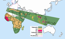
Hemoglobin E is most prevalent in mainland Southeast Asia (Thailand, Myanmar, Cambodia, Laos, Vietnam[9]), Sri Lanka, Northeast India and Bangladesh. In mainland Southeast Asia, its prevalence can reach 30 or 40%, and Northeast India, in certain areas it has carrier rates that reach 60% of the population. In Thailand the mutation can reach 50 or 70%, and it is higher in the northeast of the country. In Sri Lanka, it can reach up to 40% and affects those of Sinhalese and Vedda descent.[10][11] It is also found at high frequencies in Bangladesh and Indonesia.[12][13] The trait can also appear in people of Turkish, Chinese and Filipino descent.[1] The mutation is estimated to have arisen within the last 5,000 years.[14] In Europe, there have been found cases of families with hemoglobin E, but in these cases, the mutation differs from the one found in South-East Asia. This means that there may be different origins of the βE mutation.[15][16]
References
- ^ a b "Hemoglobin e Trait - Health Encyclopedia - University of Rochester Medical Center".
- ^ "Archived copy" (PDF). Archived from the original (PDF) on 2014-06-24. Retrieved 2017-06-08.
{{cite web}}: CS1 maint: archived copy as title (link) - PMID 13353880.
- ^ a b Bachir, D; Galacteros, F (November 2004), Hemoglobin E disease. (PDF), Orphanet Encyclopedia, retrieved January 13, 2014
- ^ Arkansas Department of Health. "Sickle-Hemoglobin E Disease Fact Sheet" (PDF).
- PMID 37076455.
- S2CID 10435042.
- ^ PMID 22908199.
- ^ Hemoglobin E Trait, University of Rochester Medical Center, retrieved January 13, 2014
- ISBN 9788120725621.
- ^ http://php.scripts.psu.edu/nxm2/1985%20Publications/1985-roychoudhury-nei.pdf [bare URL PDF]
- ISBN 9781402022319.
- PMID 22089616.
- PMID 15114532. Free full text
- ISBN 978-1-4051-4265-6.
