
Third ventricle
This article needs additional citations for verification. (November 2007) |
| Third ventricle | |
|---|---|
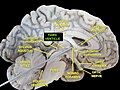
Interventricular foramina (Monro) Yellow – Third ventricle Red – Cerebral aqueduct (Sylvius) Purple – fourth ventricle Green – continuous with the central canal (Apertures to subarachnoid space are not visible) | |
| Details | |
| Identifiers | |
| Latin | ventriculus tertius cerebri |
| MeSH | D020542 |
| NeuroNames | 446 |
| NeuroLex ID | birnlex_714 |
| TA98 | A14.1.08.410 |
| TA2 | 5769 |
| FMA | 78454 |
| Anatomical terms of neuroanatomy] | |
The third ventricle is one of the four connected
Running through the third ventricle is the interthalamic adhesion, which contains thalamic neurons and fibers that may connect the two thalami.
Structure
The third ventricle is a narrow, laterally flattened, vaguely rectangular region, filled with
The lateral side of the ventricle is marked by a sulcus – the hypothalamic sulcus – from the inferior side of the interventricular foramina to the anterior side of the cerebral aqueduct. The lateral border posterior/superior of the sulcus constitutes the thalamus, while anterior/inferior of the sulcus it constitutes the hypothalamus. The interthalamic adhesion usually tunnels through the thalamic portion of the ventricle, joining together the left and right halves of the thalamus, although it is sometimes absent, or split into more than one tunnel through the ventricle; it is currently unknown whether any nerve fibres pass between the left and right thalamus via the adhesion (it has more resemblance to a herniation than a commissure).
The posterior border of the ventricle primarily constitutes the epithalamus. The superior part of the posterior border constitutes the habenular commissure, while more centrally it the pineal gland, which regulates sleep and reacts to light levels. Caudal of the pineal gland is the posterior commissure; nerve fibres reach the posterior commissure from the adjacent midbrain, but their onward connection is currently uncertain. The commissures create concavity to the shape of the posterior ventricle border, causing the suprapineal recess above the habenular, and the deeper pineal recess between the habenular and posterior commissures; the recesses being so-named due to the pineal recess being bordered by the pineal gland.

The anterior wall of the ventricle forms the
The portion of the floor immediately posterior of the optic chiasm distends inferiorly, and slightly anteriorly, to form a funnel (the
The
Development
The third ventricle, like other parts of the ventricular system of the brain, develops from the neural canal of the
The hypothalamic region of the ventricle develops from the ventral portion of the neural tube, while the thalamic region develops from the dorsal portion; the wall of the tube thickens and becomes the hypothalamus and thalamus respectively. The hypothalamic area of the ventricle begins to distend ventrally during the 5th week of development, creating the infundibulum and posterior pituitary; an outgrowth from the stomodeum (the future mouth) gradually extends towards it, to form the anterior pituitary.
The optic recess is noticeable by the end of the 6th week, by which time a bend is distinguishable in the dorsal portion of the ventricle border. Rostral of the bend, the medial dorsal portion of the ventrical begins to flatten, and become secretory (i.e. choroid plexus), forming the roof of the ventricle. Caudal of the bend, the ventricle border forms the epithalamus, and begins to distend towards the parietal bone (in lower vertebrates, it distends more specifically to the parietal eye); the border of the distention forms the pineal gland.
Clinical significance
The floor of the third ventricle is formed by hypothalamic structures and this can be opened
Several studies have found evidence of ventricular enlargement to be associated with major
A chordoid glioma is a rare tumour that can arise in the third ventricle.[7]
Additional images
-
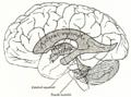 Third ventricle
 -
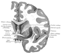
 Coronal section of brain immediately in front of pons.
 -
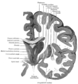 Coronal section of brain through intermediate mass of third ventricle.
 -
 Coronal section of lateral and third ventricles.
 -
 Drawing of a cast of the ventricular cavities, viewed from above.
 -
 Coronal section of brain through anterior commissure.
 -
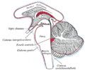 Diagram showing the positions of the three principal subarachnoid cisternæ.
 -
 Median sagittal through the hypophysis of an adult monkey. Semidiagrammatic.
 -
 Third ventricle
 -
 Third ventricle


See also
- Biology of depression
- Suprapineal recess
- Tanycytesline the bottom of the ventricle
References
- ISBN 9788131237274.
- ^
Le, Tao; Bhushan, Vikas; Vasan, Neil (2010). First Aid for the USMLE Step 1: 2010 20th Anniversary Edition. USA: ISBN 978-0-07-163340-6.
- S2CID 1913782. Retrieved September 25, 2013.
- S2CID 25819902. Retrieved September 25, 2013.
- PMID 19150053.
- PMID 16316783.
- PMID 26018908.