
Vision in fish
.jpg)
Among
Water as a visual environment


Fish and other aquatic animals live in a different light environment than terrestrial species do. Water absorbs light so that with increasing depth the amount of light available decreases quickly. The optical properties of water also lead to different wavelengths of light being absorbed to different degrees. For example, visible light of long wavelengths (e.g. red, orange) is absorbed more in water than light of shorter wavelengths (green, blue). Ultraviolet light (even shorter wavelength than violet) can penetrate deeper than visual spectra.[5] Besides these universal qualities of water, different bodies of water may absorb light of different wavelengths due to varying salt and/or chemical presence in the water.
Water is very effective at absorbing incoming light, so the amount of light penetrating the ocean declines rapidly (is attenuated) with depth. In clear ocean water, at one metre depth only 45% of the solar energy that falls on the ocean surface remains. At 10 metres depth only 16% of the light is still present, and only 1% of the original light is left at 100 metres. No light penetrates beyond 1000 metres.[6]
In addition to overall attenuation, the oceans absorb the different wavelengths of light at different rates. The wavelengths at the extreme ends of the visible spectrum are attenuated faster than those wavelengths in the middle. Longer wavelengths are absorbed first. In clear ocean waters red is absorbed in the upper 10 metres, orange by about 40 metres, and yellow disappears before 100 metres. Shorter wavelengths penetrate further, with blue and green light reaching the deepest depths.[6] This is why things appear blue underwater: how colours are perceived by the eye depends on the wavelengths of light that are received by the eye. An object appears red to the eye because it reflects red light and absorbs other colours. So the only colour reaching the eye is red. Blue is the only colour of light available at depth underwater, so it is the only colour that can be reflected back to the eye, and everything has a blue tinge under water. A red object at depth will not appear red because there is no red light available to reflect off of the object. Objects in water will only appear as their real colours near the surface where all wavelengths of light are still available, or if the other wavelengths of light are provided artificially, such as by illuminating the object with a dive light.[6]
Structure and function


Fish eyes are broadly similar to those of other vertebrates – notably the
Lenses are normally spherical but can be slightly elliptical in some species. Compared to terrestrial vertebrates, fish lenses are generally more dense and spherical. In the aquatic environment there is not a major difference in the refractive index of the cornea and the surrounding water (compared to air on land) so the lens has to do the majority of the refraction.[7] Due to "a refractive index gradient within the lens — exactly as one would expect from optical theory",[8] the spherical lenses of fish are able to form sharp images free from spherical aberration.[7]
Once light passes through the lens, it is transmitted through a transparent liquid medium until it reaches the retina, containing the photoreceptors. Like other vertebrates, the photoreceptors are on the inside layer so light must pass through layers of other neurons before it reaches them. The retina contains rod cells and cone cells.[5] There are similarities between fish eyes and those of other vertebrates. Usually, light enters through the fish eye at the cornea and passes through the pupil in order to reach the lens. Most fish species have a fixed size of the pupil while a few species have a muscular iris that allows for the adjustment of the pupil diameter.
Fish eyes have a more spherical lens than other terrestrial vertebrates. Adjustment of focus in mammals and birds is normally done by changing the shape of the eye lens while in fish this is done through moving the lens further from or closer to the retina. The retina of a fish generally has both rod cells and cone cells that are responsible for scotopic and photopic vision. Most fish species have color vision. There are some species that are capable of seeing ultraviolet while some are sensitive to polarized light.[9]
The fish retina has rod cells that provide high visual sensitivity in low light conditions and cone cells that provide higher temporal and spatial resolution than the rod cells are capable of. They allow for the possibility of color vision through the comparison of absorbance across different types of cones.[10] According to Marshall et al., most animals in the marine habitat possess no or relatively simple color vision. However, there is a greater diversity in color vision in the ocean than there is on land. This is mainly due to extremes in photic habitat and colour behaviours.[11]
The retina
Within the retina,
The distribution of photoreceptors across the retina is not uniform. Some areas have higher densities of cone cells, for example (see
Some species have a tapetum, a reflective layer which bounces light that passes through the retina back through it again. This enhances sensitivity in low light conditions, such as nocturnal and deep sea species, by giving photons a second chance to be captured by photoreceptors.[7] However this comes at a cost of reduced resolution. Some species are able to effectively turn their tapetum off in bright conditions, with a dark pigment layer covering it as needed.[5]
The retina uses a lot of oxygen compared to most other tissues, and is supplied with plentiful oxygenated blood to ensure optimal performance.[5]
Accommodation
Stabilising images
There is a need for some mechanism that
The diagram on the right shows the horizontal
- "Goldfish" shows the principal three-neuronal vestibulo-ocular reflex linking the motoneurons.[18]
- "Flatfish" shows that after 90° displacement of the vestibular relative to visual axis (metamorphosis) compensatory eye movements are produced by redirecting horizontal canal signals to vertical and oblique motoneurons.[19][20]
- In "Shark" horizontal canal/second order Ascending tract of Deiter's.[20]
Ultraviolet
Fish vision is mediated by four visual pigments that absorb various wavelengths of light. Each pigment is constructed from a chromophore and the transmembrane protein, known as opsin. Mutations in opsin have allowed for visual diversity, including variation in wavelength absorption.[21] A mutation of the opsin on the SWS-1 pigment allows some vertebrates to absorb UV light (≈360 nm), so they can see objects that reflect UV light.[22] A wide range of fish species has developed and maintained this visual trait throughout evolution, suggesting it is advantageous. UV vision may be related to foraging, communication, and mate selection.
The leading theory regarding the evolutionary selection of UV vision in select fish species is due to its strong role in mate selection. Behavioral experiments show that African cichlids utilise visual cues when choosing a mate. Their breeding sites are typically in shallow waters with high clarity and UV light penetration. Male African cichlids are largely a blue colour that is reflective in UV light. Females are able to correctly choose a mate of their species when these reflective visual cues are present. This suggests that UV light detection is crucial for correct mate selection.[23] UV reflective colour patterns also enhance male attractiveness in guppies and three-spined sticklebacks. In experimental settings, female guppies spent significantly more time inspecting males with UV-reflective colouring than those with UV reflection blocked.[24] Similarly, female three-spined sticklebacks preferred males viewed in full spectrum over those viewed in UV blocking filters.[25] These results strongly suggest the role of UV detection in sexual selection and, thus, reproductive fitness. The prominent role of UV light detection in fish mate choice has allowed the trait to be maintained over time. UV vision may also be related to foraging and other communication behaviors.
Many species of fish can see the ultraviolet end of the spectrum, beyond the violet.[26]
Ultraviolet vision is sometimes used during only part of the life cycle of a fish. For example, juvenile brown trout live in shallow water where they use ultraviolet vision to enhance their ability to detect zooplankton. As they get older, they move to deeper waters where there is little ultraviolet light.[22]
The
Polarised light
It is not easy to establish whether a fish is sensitive to
Double cones
Most fish have double cones, a pair of cone cells joined to each other. Each member of the double cone may have a different peak absorbance, and behavioural evidence supports the idea that each type of individual cone in a double cone can provide separate information (i.e. the signal from individual members of the double cone are not necessarily summed together).[32]
Adaptation to habitat

The four-eyed fish feeds at the surface of the water with eyes that allow it to see both above and below the surface at the same time.

1 Underwater retina 2) Lens 3) Air pupil
4) Tissue band 5) Iris 6) Underwater pupil
7) Air retina 8) Optic nerve
.png)
Fishes that live in surface waters down to about 200 metres,
Still deeper down the
At the very bottom of the ocean
...bony fish as a rule have a marked tendency to be flattened in a vertical direction.... It was natural, therefore, that when the ancestors of [flatfish] took to the sea bottom, they should have lain on one side.... But this raised the problem that one eye was always looking down into the sand and was effectively useless. In evolution this problem was solved by the lower eye 'moving' round to the upper side.[44]
-
![Most deep-sea fish cannot see red light. The deepwater stoplight loosejaw produces red bioluminescence so it can hunt with an effectively invisible beam of light.[45]](//upload.wikimedia.org/wikipedia/commons/thumb/b/b2/Malacosteus.JPG/179px-Malacosteus.JPG) Most deep-sea fish cannot see red light. The deepwater stoplight loosejaw produces red bioluminescence so it can hunt with an effectively invisible beam of light.[45]
Most deep-sea fish cannot see red light. The deepwater stoplight loosejaw produces red bioluminescence so it can hunt with an effectively invisible beam of light.[45] -
 When the larvae of a flatfish grows, the eye on one side rotates to the other side so the fish can rest on the seafloor.
 -
 The European plaice is a flatfish with raised eyes, so when it buries itself in sand for camouflage it can still see.
![Most deep-sea fish cannot see red light. The deepwater stoplight loosejaw produces red bioluminescence so it can hunt with an effectively invisible beam of light.[45]](/File:Malacosteus.JPG)


Prey usually have eyes on the sides of their head so they have a large field of view, from which to avoid predators. Predators usually have eyes in front of their head so they have better depth perception.[46][47] Benthic predators, like flatfish, have eyes arranged so they have a binocular view of what is above them as they lie on the bottom.
Colouration
Fish have evolved sophisticated ways of using
While these tools may be effective as predator avoidance mechanisms, they also serve as equally effective tools for the predators themselves. For example, the deepwater
-
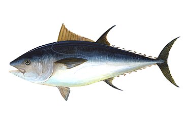 Epipelagic fish, like this Atlantic bluefin tuna, are typically countershaded with silvery colours.
 -
 The foureye butterflyfish has false eyes on its back end, confusing predators about which is the front end of the fish.
 -
 The John Dory has a large eye spot in the middle of its body, confusing prey.


Some fish species also display
The
Barreleyes


generally directed upwards, but can also be swivelled forward
---------------------------------------------------------------------
Right: The brownsnout spookfish is the only vertebrate known
to employ a mirror eye (as well as a lens):
(1) diverticulum (2) main eye
(a) retina (b) reflective crystals (c) lens (d) retina
| External videos | |
|---|---|
Barreleyes are a family of small, unusual-looking mesopelagic fishes, named for their barrel-shaped, tubular eyes which are generally directed upwards to detect the silhouettes of available prey.[53][54] Barreleyes have large, telescoping eyes which dominate and protrude from the skull. These eyes generally gaze upwards, but can also be swivelled forwards in some species. Their eyes have a large lens and a retina with an exceptional number of rod cells and a high density of rhodopsin (the "visual purple" pigment); there are no cone cells.[53]
The barreleye species,
Another barreleye species, the
Sharks

Shark
Other examples

Small fish often
Fish are normally cold-blooded, with body temperatures the same as the surrounding water. However, some oceanic predatory fish, such as swordfish and some shark and tuna species, can warm parts of their body when they hunt for prey in deep and cold water. The highly visual swordfish uses a heating system involving its muscles which raises the temperature in its eyes and brain by up to 15 °C. The warming of the retina improves the rate at which the eyes respond to changes in rapid motion made by its prey by as much as ten times.[66][67][68]
Some fish have
Many species of
Distance sensory systems
Visual systems are distance sensory systems which provide fish with data about location or objects at a distance without a need for the fish to directly touch them. Such distance sensing systems are important, because they allow communication with other fish, and provide information about the location of food and predators, and about avoiding obstacles or maintaining position in fish schools. For example, some schooling species have "schooling marks" on their sides, such as visually prominent stripes which provide reference marks and help adjacent fish judge their relative positions.[73] But the visual system is not the only one that can perform such functions. Some schooling fish also have a lateral line running the length of their bodies. This lateral line enables the fish to sense changes in water pressure and turbulence adjacent to its body. Using this information, schooling fish can adjust their distance from adjacent fish if they come too close or stray too far.[73]
The visual system in fish is augmented by other sensing systems with comparable or complementary functions. Some fish are blind, and must rely entirely on alternate sensing systems.
See also
- Arthropod eye
- Matthiessen's ratio
- Mollusc eye
- Parietal eye
- Simple eye in invertebrates
- Visual system
Notes
- S2CID 22940203.
- PMID 18026166. See also Lamb et al.'s The origin of the Vertebrate Eye, 2008.
- PMID 18026166.
- ^ Ocean Explorer NOAA. Updated: 26 August 2010.
- ^ a b c d e f g h i j k Helfman et al. 2009, pp. 84–87.
- ^ a b c Webb, Paul (2019) Introduction to Oceanography, chapter 6.5 Light, Rebus Community, Roger Williams University, open textbook.
 Material was copied from this source, which is available under a Creative Commons Attribution 4.0 International License.
- ^ ISBN 9780199581146.
- S2CID 4408533.
- ^ PMID 30700806.
- PMID 27218707.
- S2CID 20978931.
- S2CID 19066809.
- PMID 16572506.
- ISBN 9780195369748.
- S2CID 17757889.
- ISBN 978-81-7075-029-1.
- PMID 311828.
- S2CID 13004673.
- PMID 4067626.
- ^ PMID 11535684.
- .
- ^ PMID 12824471.
- S2CID 5420659.
- S2CID 53172856.
- S2CID 937644.
- .
- S2CID 140204848.
- S2CID 3743161.
- ^ Horváth G and Varjú D (2004)
Polarized light in animal vision: polarization patterns in nature p. 294, Springer. ISBN 978-3-540-40457-6.
- Denton, EJ; Nichol, JAC (1965). "Polarization of light reflected from the silvery exterior of the bleak Alburnus alburnus"(PDF). J. Mar. Biol. Assoc. U. K. 150: 78–94.
- PMC 1691948.
- PMID 20129950.
- ^ Froese, Rainer; Pauly, Daniel (eds.) (2010). "Gigantura chuni" in FishBase. October 2010 version.
- ^ Froese, Rainer; Pauly, Daniel (eds.) (2009). "Dissostichus mawsoni" in FishBase. August 2009 version.
- ISBN 978-0-471-25031-9.)
{{cite book}}: CS1 maint: multiple names: authors list (link - ^ Froese, Rainer; Pauly, Daniel (eds.) (2007). "Anableps anableps" in FishBase. Mar 2007 version.
- ^ Moyle & Cech 2004, p. 585.
- S2CID 83905458.
- PMID 841297.
- ISBN 978-0-12-547665-2.
- ^ Ryan P "Deep-sea creatures: The bathypelagic zone" Te Ara – the Encyclopedia of New Zealand. Updated 21 September 2007.
- ^ Moyle & Cech 2004, p. 587.
- ISBN 978-0-12-547665-2.
- ISBN 978-0-14-014481-9.
- S2CID 1038874.
- ^ "Carnivores". U.S. Department of the Interior, Bureau of Land Management. 14 December 2009. Archived from the original on 14 June 2011. Retrieved 28 March 2011.
- Stanford. Archived from the originalon 5 July 2010. Retrieved 11 May 2010.
- ^ Countershading BBC: Science and Nature. Retrieved 28 September 2011.
- ^ Fishy friends and fishy foes Preparation manual, Long Beach Marine Institute.
- .
- ^ FishBaseFroese, Rainer; Pauly, Daniel (eds.) (2009). "Chaetodon capistratus" in FishBase. July 2009 version.
- ^ Walrond, Carl (2006) Coastal fish - Fish of the open sea floor, Te Ara: Encyclopedia of New Zealand. Accessed 28 May 2019.
- ^ S2CID 85768623.
- ^ ProQuest 207224476.
- "Researchers solve mystery of deep-sea fish with tubular eyes and transparent head". Monterey Bay Aquarium Research Institute (Press release). 23 February 2009.
- ^ Froese, Rainer; Pauly, Daniel (eds.) (2011). "Macropinna microstoma" in FishBase. September 2011 version.
- ^ S2CID 18680315.
- ^ Reporter, Lewis Smith, Environment (8 January 2009). "Fish with four eyes can see through the deep sea gloom". The Times.
{{cite news}}: CS1 maint: multiple names: authors list (link) - ^ Martin, R. Aidan. "Vision and a Carpet of Light". ReefQuest Centre for Shark Research. Retrieved 22 August 2009.
- S2CID 30148811.
- "Sharks are colour-blind, new study finds". Australian Geographic. 19 January 2011.
- ^ Gill, Victoria (18 January 2011). "Sharks are probably colour-blind". BBC News. Retrieved 19 January 2011.
- S2CID 30148811.
- S2CID 4184043.
- ^ Moyle & Cech 2004, p. [page needed].
- S2CID 53202810.
- S2CID 53205760.
- S2CID 14070646.
- .
- ^ Helfman et al. 2009, pp. 95–97.
- .
- ^ Johnson JA and Esser R (2009) "http://www.fishculturesection.org/Aquanotes/pdf/Aq_App_Note_1_April_2009.pdf Walleye Culture – Habituation to Feed in the Dark" American Fisheries Society, Aquaculture Application Note.
- ^ PMID 12364396.
- PMID 23270452.
- ^ a b Bone & Moore 2008, pp. 418–422.
- ^ Bone & Moore 2008, p. 311.
- ISBN 978-971-02-0003-0.
- PMID 17904741.
- PMID 19760113.
- S2CID 13996233.
References
- Bone, Quentin; Moore, Richard (2008). Biology of Fishes. Garland Science. ISBN 978-0-203-88522-2.
- Helfman, Gene; Collette, Bruce B.; Facey, Douglas E.; Bowen, Brian W. (2009). The Diversity of Fishes: Biology, Evolution, and Ecology. John Wiley & Sons. ISBN 978-1-4443-1190-7.
- Moyle, Peter B.; Cech, Joseph J. (2004). Fishes: An Introduction to Ichthyology. Pearson Prentice Hall. ISBN 978-0-13-100847-2.
Further reading
- Arthur, Joseph; Nicol, Colin; Somiya, Hiroaki (1989). The eyes of fishes. Clarendon Press. ISBN 978-0-19-857195-7.
- Douglas, R. H. & Djamgoz, M. (eds) (1990) The Visual System of Fish. Chapman and Hall, 526 pp.
- Lamb, Trevor D. (14 June 2011). "Evolution of the Eye". Scientific American. 305 (1): 64–69. .
- Land, Michael F and Nilsson, Dan-Eric (2012) Animal Eyes Oxford University Press. ISBN 9780199581146.
- Lamb, Trevor D.; Collin, Shaun P.; Pugh, Edward N. (December 2007). "Evolution of the vertebrate eye: opsins, photoreceptors, retina and eye cup". Nature Reviews. Neuroscience. 8 (12): 960–976. PMID 18026166.
- "Keeping an eye on evolution". Phys.org. 3 December 2007.
- Nilsson, DE; Pelger, S (22 April 1994). "A pessimistic estimate of the time required for an eye to evolve". Proceedings of the Royal Society of London. Series B: Biological Sciences. 256 (1345): 53–58. S2CID 13061351.
- Berlinski, David (2002) Has Darwin Met His Match? Page 34, The Vexing Eye (Letter). Commentary, 1 December 2002.
- Nilsson, Dan-E. "Beware of Pseudo-science: a response to David Berlinski's attack on my calculation of how long it takes for an eye to evolve". Talk Reason.
- Meyer-Rochow, Victor Benno; Coddington, Paul Edward (2003). "Eyes and vision of the New Zealand torrentfish Cheimarrichthys foster VON HAAST (1874): Histology, photochemistry and electrophysiology". In Val, Adalberto Luís; Kapoor, B. G. (eds.). Fish Adaptations. Science Publishers. pp. 337–383. ISBN 978-1-57808-249-0.
- "Evolution of the Eye" – video on Nilsson-Pelger model (scroll down)
Bibliography
- Marshall, Justin; Carleton, Karen L; Cronin, Thomas (October 2015). "Colour vision in marine organisms". Current Opinion in Neurobiology. 34: 86–94. S2CID 20978931.
- Kamijo, Makiko; Kawamura, Mayuko; Fukamachi, Shoji (May 2018). "Loss of red opsin genes relaxes sexual isolation between skin-colour variants of medaka". Behavioural Processes. 150: 25–28. S2CID 4239046.
External links
Vision in animals | ||
|---|---|---|
| Vision | | |
| Eyes |
| |
| Evolution | ||
| Coloration | ||
| Related topics | ||