
Chorion
| Chorion | |
|---|---|
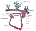
 Diagram showing the chorion of a chicken egg | |
 Human fetus enclosed in the amnion | |
| Details | |
| Identifiers | |
| Latin | chorion |
| TE | E5.11.3.1.1.0.3 |
| Anatomical terminology | |
The chorion is the outermost
Structure
In humans and other
Layers
The chorion consists of two layers: an outer formed by the trophoblast, and an inner formed by the extra-embryonic mesoderm.
The trophoblast is made up of an internal layer of cubical or prismatic cells, the
Growth
The chorion undergoes rapid proliferation and forms numerous
The chorionic villi are at first small and non-vascular, and consist of the trophoblast only, but they increase in size and
Blood is carried to the villi by the paired
Parts

The part of the chorion that is in contact with the decidua capsularis undergoes atrophy, so that by the fourth month scarcely a trace of the villi is left. This part of the chorion becomes smooth,[2] and is named the chorion laeve (from the Latin word levis, meaning smooth). As it takes no share in the formation of the placenta, this is also named the non-placental part of the chorion. As the chorion grows, the chorion laeve comes in contact with the decidua parietalis and these layers fuse.
The villi at the embryonic pole, which is in contact with the
Thus the placenta develops from the chorion frondosum and the decidua basalis.
Monochorionic twins
Monochorionic twins are
Infections
Recent studies indicate that the chorion may be susceptible to
Other animals

In reptiles, birds, and monotremes, the chorion is one of the four extraembryonic membranes that make up the amniotic egg that provide for the nutrients and protection needed for the embryo's survival. It is located inside the albumen, which is the white of the egg. It encloses the embryo and the rest of the embryonic system. The chorion is also present in insects. During growth and development of the embryo, there is an increased need for oxygen. To compensate for this, the chorion and the allantois fuse together to form the chorioallantoic membrane. Together these form a double membrane, which functions to remove carbon dioxide and to replenish oxygen through the porous shell. At the time of hatching, the fetus becomes detached from the chorion as it emerges from the shell.
In
Additional images
-
 Section through the embryo.
 -
 Diagram of the human embryo.
 -
 Diagram illustrating early formation of allantois and differentiation of body-stalk.
 -
 Diagram showing later stage of allantoic development with commencing constriction of the yolk-sac.
 -
 Scheme of placental circulation.


See also
- Choriogenesis
- Chorioamnionitis, an inflammation of the chorion and amnion, usually due to bacterial infection
- Chorionic hematoma
- trophoblasts, including choriocarcinoma, a highly invasive cancer.
References
![]() This article incorporates text in the public domain from page 60 of the 20th edition of Gray's Anatomy (1918)
This article incorporates text in the public domain from page 60 of the 20th edition of Gray's Anatomy (1918)
- ISBN 978-0-12-815145-7.
- ^ PMID 26003500.
- PMID 16281049.
- ^ ISBN 0-8247-2844-0.
- PMID 30078192.
- PMID 30078192.
- S2CID 53106591.
- PMID 31549405.
- S2CID 53106591.
- ^ Chapman, R.F. (1998) "The insects: structure and function", Section The egg and embryology. Previewed in Google Books [1] on 26 Sep 2009.
- ^ “The Octopoda are characterized by eggs that have only a chorion as an envelope”https://www.sciencedirect.com/topics/agricultural-and-biological-sciences/octopoda
External links
- Histology image: 19903loa – Histology Learning System at Boston University — "Female Reproductive System: placenta, chorionic plate"
- McGill