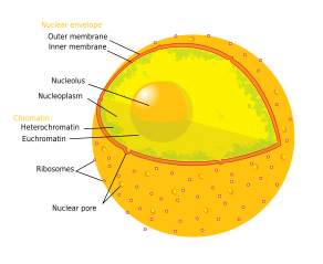
Nucleolus

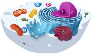
Animal cell diagram | |
|---|---|
 Components of a typical animal cell:
|
The nucleolus (
History
The nucleolus was identified by bright-field microscopy during the 1830s.[7] Theodor Schwann in his 1939 treatise describes that Schleiden had identified small corpuscleus in nuclei, and names the structure "Kernkörperchen". In a 1947 translation of the work to English, the structure receives the name of "nucleolus".[8][9]
In addition to these peculiarities of the cytoblast, already made known by Brown and Meyen, Schleiden has discovered in its interior a small corpuscle (see plate I, fig. 1, 4,) which, in the fully-developed cytoblast, looks like a thick ring, or a thick-walled hollow globule. It appears, however, to present a different appearance in different cytoblasts. Sometimes only the external sharply-defined circle of this ring can be distinguished, with a dark point in the centre,—occasionally, and indeed most frequently, only a sharply circumscribed spot. In other instances this spot is very small, and sometimes cannot be recognized at all. As it will frequently be necessary to speak of this body in the following treatise, I will for brevity’s sake name it the “nucleolus,” (Kernkorperchen, ‘nucleus-corpuscle.”)
— Theodor Schwann, translated by Henry Smith, Microscopical Researches Into the Accordance in the Structure and Growth of Animals and Plants, page 3
Little was known about the function of the nucleolus until 1964, when a study
Structure
Three major components of the nucleolus are recognized: the fibrillar center (FC), the dense fibrillar component (DFC), and the granular component (GC).[1] Transcription of the rDNA occurs in the FC.[13] The DFC contains the protein fibrillarin,[13] which is important in rRNA processing. The GC contains the protein nucleophosmin,[13] (B23 in the external image), which is also involved in ribosome biogenesis.
However, it has been proposed that this particular organization is only observed in higher eukaryotes and that it evolved from a bipartite organization with the transition from anamniotes to amniotes. Reflecting the substantial increase in the DNA intergenic region, an original fibrillar component would have separated into the FC and the DFC.[14]
Another structure identified within many nucleoli (particularly in plants) is a clear area in the center of the structure referred to as a nucleolar vacuole.[15] Nucleoli of various plant species have been shown to have very high concentrations of iron[16] in contrast to human and animal cell nucleoli.
The nucleolus ultrastructure can be seen through an electron microscope, while the organization and dynamics can be studied through fluorescent protein tagging and fluorescent recovery after photobleaching (FRAP). Antibodies against the PAF49 protein can also be used as a marker for the nucleolus in immunofluorescence experiments.[17]
Although usually only one or two nucleoli can be seen, a diploid human cell has ten nucleolus organizer regions (NORs) and could have more nucleoli. Most often multiple NORs participate in each nucleolus.[18]
Function and ribosome assembly

In
Transcription of rRNA yields a long precursor molecule (45S pre-rRNA), which still contains the ITS and ETS. Further processing is needed to generate the 18S RNA, 5.8S, and 28S RNA molecules. In eukaryotes, the RNA-modifying enzymes are brought to their respective
In higher
In human endometrial cells, a network of nucleolar channels is sometimes formed. The origin and function of this network have not yet been clearly identified.[22]
Sequestration of proteins
In addition to its role in ribosomal biogenesis, the nucleolus is known to capture and immobilize proteins, a process known as nucleolar detention. Proteins that are
See also
References
- ^ PMID 25436580.
- ISBN 978-0-470-01617-6.
- PMID 24631655.
- PMID 35096082.
- PMID 24389329.
- PMID 25464032.
- PMID 21106648.
- ^ "Mikroskopische Untersuchungen über die Uebereinstimmung in der Struktur und dem Wachsthum der Thiere und Pflanzen / Von Th. Schwann. Mit vier Kupfertafeln". Wellcome Collection. Retrieved 12 March 2024.
- ^ Schwann, Theodor; Schwann, Theodor; Smith, Henry; Schleiden, M. J. (1847). Microscopical researches into the accordance in the structure and growth of animals and plants. London: Sydenham Society. p. 3.
- PMID 14106673.
- PMID 5963987.
- PMID 5943882.
- ^ PMID 18046571.
- PMID 8799814.
- PMID 21719700.
- ^ PAF49 antibody | GeneTex Inc. Genetex.com. Retrieved 2019-07-18.
- ^ von Knebel Doeberitz M, Wentzensen N (2008). "The Cell: Basic Structure and Function". Comprehensive Cytopathology (third ed.).
- ISBN 978-0-7817-2265-0.
- ISBN 978-0-8153-3218-3.
- ISBN 978-0-87893-220-7.
- .
- PMID 22284675.
Further reading
- Cooper GM (2000). "The Nucleolus". The Cell: A Molecular Approach (2nd ed.). Sunderland MA: Sinauer Associates. ISBN 978-0-87893-106-4.
- Tiku V, Antebi A (August 2018). "Nucleolar Function in Lifespan Regulation". Trends in Cell Biology. 28 (8): 662–672. S2CID 29167518.
- JoAnna Klein (20 May 2018). "The Thing Inside Your Cells That Might Determine How Long You Live". The New York Times.
External links
- Nucleolus under electron microscope II at uni-mainz.de
- Nuclear Protein Database – search under compartment
- Cell+Nucleolus at the U.S. National Library of Medicine Medical Subject Headings (MeSH)
- Histology image: 20104loa – Histology Learning System at Boston University
