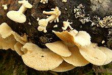
Panellus stipticus
| Panellus stipticus | |
|---|---|

| |
| Scientific classification | |
| Domain: | Eukaryota |
| Kingdom: | Fungi |
| Division: | Basidiomycota |
| Class: | Agaricomycetes |
| Order: | Agaricales |
| Family: | Mycenaceae |
| Genus: | Panellus |
| Species: | P. stipticus
|
| Binomial name | |
| Panellus stipticus | |
| Synonyms | |
| |
| Panellus stipticus | |
|---|---|
| Gills on hymenium | |
| Cap is convex or offset | |
saprotrophic | |
| Edibility is inedible | |
Panellus stipticus, commonly known as the bitter oyster, the astringent panus, the luminescent panellus, or the stiptic fungus, is a species of
Panellus stipticus is one of several dozen species of fungi that are
Taxonomy and phylogeny
| ||||||||||||||||||||||||
| Phylogeny and relationships of P. stipticus and related species based on ribosomal DNA sequences[1] |
The species was first named Agaricus stypticus by the French botanist
Panellus stipticus is the
The fungus is
Description

The fungus normally exists unseen, in the form of a mass of threadlike
Microscopic features
Various microscopic characteristics may be used to help identify the fungus from other morphologically similar species. A spore print of P. stipticus, made by depositing a large number of spores in a small area, reveals their color to be white.[21] Viewed with a microscope, the spores are smooth-walled, elliptical to nearly allantoid (sausage-shaped), with dimensions of 3–6 by 2–3 µm. Spores are amyloid, meaning that they will absorb iodine and become bluish-black when stained with Melzer's reagent,[30] but this staining reaction has been described as "relatively weak".[1]
The
The flesh of the cap consists of a number of microscopically distinct layers of tissue. The cuticle of the cap (known as the pileipellis) is between 8–10 µm thick,[32] and is made of a loose textura intricata, a type of tissue in which the hyphae are irregularly interwoven with distinct spaces between them.[15] The cuticle hyphae are thick-walled to thin-walled, with scattered inconspicuous cystidia measuring 40–55 by 3.5–5.5 µm. These cystidia located in the cap (pileocystidia) are cylindrical, thin-walled, yellow in Melzer's reagent, hyaline in KOH, sometimes with amorphous dingy brown material coating the walls. Beneath the cuticle layer is a zone 54–65 µm thick, made of very loosely entwined, thin-walled hyphae, 2–3 µm in thickness, with clamps at the septa. Below this is a zone 208–221 µm in thickness, in which the densely compacted hyphae, 3–8 µm in diameter, have swollen, gelatinized walls, and often more or less a vertical orientation. This in turn is followed by a layer 520 µm in thickness, formed of loosely interwoven hyphae, 2–8 µm in width, some of which have thin walls with clamps at the septa, whilst others have somewhat thickened gelatinized walls.[32] The flesh of the cap has a layer of upright hyphae bending into a lower layer of interwoven hyphae with diameters of 2.5–8 µm. The flesh of the gills is similar to that of the lower cap.[15]
Similar species
Species of Crepidotus having a similar shape can be distinguished by their brown spore print, compared with the white spore print of P. stipticus.[20] Schizophyllum commune has a densely hairy white to grayish cap and longitudinally split gill-folds on the underside.[25] The ruddy panus mushroom (Panus rudis) is larger, has a reddish-brown cap that fades to pinkish-tan, and shows lilac tinges when young, fresh, and moist.[23] Some Paxillus species may have a similar appearance, but they have yellow-brown spore prints.
Uses
Panellus stipticus is considered too small and bitter to be
Fruit body development
Fruit bodies first appear as tiny white knobs less than a cubic millimeter in size. In a day or two the knobs grow into a horizontal pyramidal mass, increasing in height as the hyphae lengthen. This is soon followed by the formation of a minute cap, and lengthening of the stem. The stem is about 1 mm long when the cap first begins to form. The hyphae that comprise the stem gradually cease to grow at their ends, and then start to branch, with many of the branches growing in a horizontal direction. This growth, indicated by the flattening and broadening out of the top of the stem, gives rise to the cap. The horizontally aligned hyphae grow vertical branches which remain more or less parallel, ultimately forming the dorsal tissue of the cap. Other similar downward-growing branches form the fertile hymenium, which can be seen when the cap is about 2 mm in diameter.[36]

The young cap is spherical and its growth is at first epinastic, its margin being curved inwards and pressed against the stem. In this way, the hymenium begins its development within a special enclosed chamber. As the hymenial surface increases and keeps pace with the growth of the dorsal tissue of the cap, the latter expands and exposes the gills. The gills are formed by the continual downward growth of some of the hyphae. The gills are exposed before the cap is completely developed, and before the spores are mature. Spores can be produced by fruit bodies as small as 1.3 cm (0.5 in) broad, and liberation of the spores continues until the fruit body is fully grown—a period of one month to three months, depending on the conditions of temperature and moisture. The mature spores are disseminated by the wind. When the fruit body is nearing maturity, some of the terminal portions of the hyphae of the dorsal surface of the cap separate, and as a consequence, the upper surface of the fruit body becomes granular in appearance.[36]
The fruit body projects out horizontally from the growing surface. If the position of a log is altered after young fruit bodies with the beginnings of gills have appeared, the stems of these attempt to readjust themselves in order to place the cap in a horizontal position. The cap are sometimes zonate (marked with concentric lines that form alternating pale and darker zones); this depends on changes in the humidity of the environment, as variations in the amount of moisture will cause alternating periods of acceleration or slowing of growth.[36]
A yellowish-brown pigment is diffused through the
Distribution, habitat, and ecology

Panellus stipticus is common in northern
Panellus stipticus is a
The fruit bodies are frequently attacked by slugs, which may be important agents in the dispersal of its spores.[23] White-tailed deer are also known to consume the fungus.[49]
Mating studies
Panellus stipticus uses a
Bioluminescence

An early record of luminescence noted in P. stypticus was made by the American naturalist
By careful examination, the luminosity was found to proceed from the gills and not the stipe, nor from any fragment of rotten wood attached to the specimen. This phosphorescence was not observed in all specimens brought in for examination, and seemed to depend on some peculiar condition of the air, having been noticed only in specimens gathered in damp weather or just before a storm.[56]
Canadian mycologist Buller in 1924 described the gills of P. stipticus in North America as luminescent, and noted that the fungus glows most strongly at the time of spore maturation.[42] Bioluminescence has not been observed in European specimens,[43] in Pacific North American collections, nor in strains collected from New Zealand, Russia, and Japan.[34] Although a number of reports have confirmed that eastern North American strains are luminescent,[34][44][51][57][58] non-luminescent North American strains are also known.[54] In general, the intensity of fungal bioluminescence decreases after exposure to certain contaminants; this sensitivity is being investigated as a means to develop bioluminescence-based biosensors to test the toxicity of polluted soils.[59] Most known luminescent fungi are in the genus Mycena or closely allied genera; this grouping of fungi—known as the "mycenoid lineage"—includes P. stipticus and three other Panellus species.[60]
Mycelia
The
Fruit bodies
Bioluminescent tissue in the mature fruit body is restricted to the edge of the gills (as well as the cross-veins that connect them), the junction of the gills with the stem, and the inrolled cap edge. Distribution of bioluminescence along the gill edge corresponds to the position of the cheilocystidia. Less than 10% of the light emitted from both the young and mature fruit bodies is from other tissues, including the fertile hymenial area and the stem. Fruit body luminescence is highly variable between fruit bodies found on different logs in different environments.[58]
Genetics

Using techniques of genetic complementation, Macrae paired nonluminescent monocaryons with luminescent ones, and concluded that luminosity in P. stipticus is an inherited character, and governed by a single pair of alleles in which luminosity was dominant over nonluminosity. Luminosity factors were independent of intersterility factors. In 1992, Lingle and colleagues agreed with Macrae about luminosity and stated that at least three different gene mutations could lead to the loss of luminescence. They also reported that the maximum bioluminescence was found at 525 nm, and shifted to 528 nm in deeply pigmented fruit bodies.[44]
After intercontinental compatibility tests, Petersen and Bermudes suggested that bioluminescence and compatibility were independent since bioluminescence seemed to be geographically restricted. This suggested that the ability or potential to interbreed must have been preserved since separation of P. stypticus into geographically isolated areas.[34][51]
Function
Several authors have suggested that the purpose of fungal bioluminescence is to attract
Chemical basis

In general, bioluminescence is caused by the action of
Bioremediation
As a white-rot fungus, Panellus stipticus contains enzymes that are able to break down
See also
References
- ^ a b c Jin J, Petersen RH (2001). "Phylogenetic relationships of Panellus (Agaricales) and related species based on morphology and ribosomal large subunit DNA sequences". Mycotaxon. 79: 7–21.
- ^ Bulliard JBF. (1783). Herbier de la France. Vol. 3 (in French). pp. 97–144. Archived from the original on 2010-02-12.
- ^ Fries EM. (1821). Systema Mycologicum I (in Latin). Lundin, Sweden: ex officina Berlingiana. p. 188. Archived from the original on 2010-02-12.
- ^ Fries EM. (1838). Epicrisis Systematis Mycologici (in Latin). Uppsala, Sweden: Typographia Academica. p. 399.
- ^ Gmelin JF (1792). Systema Naturae per regna tria naturae: secundum classes, ordines, genera, species, cum characteribus, differentiis, synonymis, locis (in Latin). Vol. 2 (13th ed.). Leipzig, Germany: Impensis Georg. Emanuel. Beer. p. 1411.
- ^ Kuntze O. (1898). Revisio Generum Plantarum (in Latin). Vol. 3. Leipzig: Arthur Felix. p. 506.
- ^ Wallroth CFW. (1833). Flora Cryptogamica Germaniae (in Latin). Vol. 2. Nuremberg, Germany: J.L. Schrag. p. 742.
- ^ Gray SF. (1821). A Natural Arrangement of British Plants. London, UK: Baldwin, Cradock, and Joy. p. 616.
- ^ Kummer P. (1871). Der Führer in die Pilzkunde (in German). Zerbst, Germany: E. Luppe Staude. p. 105.
- ^ Schröter J. (1885). Kryptogamen-Flora von Schlesien (in German). Vol. 3–1(1). Breslau, Poland: J.U. Kern's Verlag. p. 554.
- ^ "Panellus stipticus – Species synonymy". Index Fungorum. CAB International. Archived from the original on 2012-10-04. Retrieved 2009-12-29.
- ^ Karsten P. (1879). "Rysslands, Finlands och den Skandinaviska Halföns Hattsvampar. Förra Delen: Skifsvampar". Bidrag till Kännedom of Finlands Natur Folk (in Swedish). 32: 96–7.
- ^ a b Kuo M. (April 2007). "Panellus stipticus". MushroomExpert.Com. Retrieved 2010-01-01.
- ^ ISBN 978-3-87429-254-2.
- ^ a b c d e f g Bursdall HH Jr, Miller OK Jr (1975). "A reevaluation of Panellus and Dictyopanus (Agaricales)". Nova Hedwigia. 51: 79–91.
- ISBN 978-0-85199-377-5.
- ^ "Panellus stipticus – Names Record". Index Fungorum. CAB International. Retrieved 2009-12-29.
- ISBN 978-0-85199-826-8.
- PMID 12099793.
- ^ ISBN 978-0-395-91090-0.
- ^ ISBN 978-0-02-063690-8.
- ISBN 978-0-271-02891-0.
- ^ ISBN 978-0-442-21998-7.
- ISBN 978-0-8156-0388-7.
- ^ ISBN 978-0-8131-9039-6.
- ^ "styptic". Oxford English Dictionary online. Oxford University Press. Retrieved 2010-01-27.
- JSTOR 3752832.
- ^ ISBN 978-0-7627-3109-1.
- ISBN 978-90-5410-616-6.
- ISBN 978-0-7112-2378-3.
- ^ ISBN 978-962-201-556-2.
- ^ JSTOR 4113780.
- ISBN 978-0-520-03656-7.
- ^ JSTOR 3760252.
- ISBN 978-1-884360-01-5.
- ^ .
- ISBN 978-0-86788-063-2.
- .
- JSTOR 4115687.
- ^ JSTOR 3761652.
- ^ Miller OK Jr. (1970). "The genus Panellus in North America". Michigan Botanist. 9: 17–30.
- ^ a b Buller AHR. (1924). "The bioluminescence of Panus stipticus". Researches on Fungi. Vol. III. London, UK: Longmans, Green and Company. pp. 357–431.
- ^ .
- ^ JSTOR 3760407.
- ^ ISBN 978-0-947643-02-7.
- ISSN 0010-0730.
- ISBN 978-0-89815-169-5.
- .
- JSTOR 3795797.
- S2CID 4064765.
- ^ a b c Petersen RH, Bermudes D (1992). "Intercontinental compatibility in Panellus stypticus with a note on bioluminescence". Persoonia. 14: 457–63.
- ^ Petersen RH, Hughes KW (2003). "Phylogeographic examples of Asian biodiversity in mushrooms and their relatives" (PDF). Fungal Diversity. 13: 95–109.
- PMID 19789811.
- ^ .
- ^ PMID 17610297.
- JSTOR 3752395.
- JSTOR 3760051.
- ^ JSTOR 3760050.
- PMID 12123478.
- PMID 18264584.
- ^ JSTOR 3759900.
- PMID 13681827.
- JSTOR 3756571.
- S2CID 21001720.
- PMID 11520609.
- ^ Ewart AJ. (1906). "Note on the phosphorescence of Agaricus (Pleurotus) candescens". Victorian Naturalist. 13: 174.
- JSTOR 3493838.
- .
- ISBN 978-0-471-52229-4.
- PMID 8372704.
- .
- ISSN 0031-8655.
- ISBN 978-0-85404-136-7.
- .
- S2CID 10671228.
- PMID 12167540.
External links


