
Apex beat
This article has multiple issues. Please help improve it or discuss these issues on the talk page. (Learn how and when to remove these template messages)
|
The apex beat (lat. ictus cordis), also called the apical impulse,
Identification
The normal apex beat can be
In children the apex beat occurs in the fourth rib interspace medial to the nipple. The apex beat may also be found at abnormal locations; in many cases of dextrocardia, the apex beat may be felt on the right side.
Interpretation
-
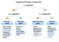 Algorithm for classification of the apex beat characters

Lateral and/or inferior displacement of the apex beat usually indicates enlargement of the heart, called cardiomegaly. The apex beat may also be displaced by other conditions:
- Pleural or pulmonary diseases
- Deformities of the chest wall or the thoracic vertebrae
Sometimes, the apex beat may not be palpable, either due to a thick chest wall, or conditions where the stroke volume is reduced; such as during ventricular tachycardia or shock.
The character of the apex beat may provide vital diagnostic clues:
- A forceful impulse indicates volume overload in the heart (as might occur in aortic regurgitation)
- An uncoordinated (ventriculardysfunction; such as an aneurysm following myocardial infarction
- A arrhythmias, such as premature ventricular contraction or atrial fibrillation.
Sustained apex beat, namely prolonged upward cardiac force during systole in a physical exam, can be seen in some chronic conditions such as hypertension and aortic stenosis, especially in elderly and females.[3]
An algorithm for the classification of some common apex beat characteristics is shown in the image
References
- ISBN 978-0-7817-8058-2.
- ^ Visualization of the point of maximal impulse and "S4" on echocardiogram: an observation. Conn Med. 2007 Feb;71(2):85–8.
- ^ "webcampus.drexelmed.edu". Archived from the original on 18 October 2018. Retrieved 17 October 2018.