Catenin beta-1
Ensembl | |||||||||
|---|---|---|---|---|---|---|---|---|---|
| UniProt | |||||||||
| RefSeq (mRNA) | |||||||||
| RefSeq (protein) | |||||||||
| Location (UCSC) | Chr 3: 41.19 – 41.26 Mb | Chr 9: 120.76 – 120.79 Mb | |||||||
| PubMed search | [3] | [4] | |||||||
| View/Edit Human | View/Edit Mouse |
Catenin beta-1, also known as β-catenin (beta-catenin), is a protein that in humans is encoded by the CTNNB1 gene.
β-Catenin is a dual function
Mutations and overexpression of β-catenin are associated with many cancers, including
Discovery
β-Catenin was initially discovered in the early 1990s as a component of a mammalian cell adhesion complex: a protein responsible for cytoplasmatic anchoring of cadherins.[11] But very soon, it was realized that the Drosophila protein armadillo – implicated in mediating the morphogenic effects of Wingless/Wnt – is homologous to the mammalian β-catenin, not just in structure but also in function.[12] Thus, β-catenin became one of the first examples of moonlighting: a protein performing more than one radically different cellular function.
Structure
Protein structure
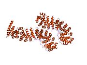
The core of β-catenin consists of several very characteristic

The segments N-terminal and far C-terminal to the ARM domain do not adopt any structure in solution by themselves. Yet these
Partners binding to the armadillo domain

As sketched above, the
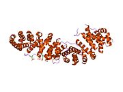
The structure of β-catenin in complex with the catenin binding domain of the transcriptional transactivation partner TCF provided the initial structural roadmap of how many binding partners of β-catenin may form interactions.[21] This structure demonstrated how the otherwise disordered N-terminus of TCF adapted what appeared to be a rigid conformation, with the binding motif spanning many beta-catenin repeats. Relatively strong charged interaction "hot spots" were defined (predicted, and later verified, to be conserved for the β-catenin/E-cadherin interaction), as well as hydrophobic regions deemed important in the overall mode of binding and as potential therapeutic small molecule inhibitor targets against certain cancer forms. Furthermore, following studies demonstrated another peculiar characteristic, plasticity in the binding of the TCF N-terminus to beta-catenin.[22][23]
Similarly, we find the familiar E-cadherin, whose cytoplasmatic tail contacts the ARM domain in the same canonical fashion.[24] The scaffold protein axin (two closely related paralogs, axin 1 and axin 2) contains a similar interaction motif on its long, disordered middle segment.[25] Although one molecule of axin only contains a single β-catenin recruitment motif, its partner the adenomatous polyposis coli (APC) protein contains 11 such motifs in tandem arrangement per protomer, thus capable to interact with several β-catenin molecules at once.[26] Since the surface of the ARM domain can typically accommodate only one peptide motif at any given time, all these proteins compete for the same cellular pool of β-catenin molecules. This competition is the key to understand how the Wnt signaling pathway works.
However, this "main" binding site on the ARM domain β-catenin is by no means the only one. The first helices of the ARM domain form an additional, special protein-protein interaction pocket: This can accommodate a helix-forming linear motif found in the coactivator BCL9 (or the closely related BCL9L) – an important protein involved in Wnt signaling.[27] Although the precise details are much less clear, it appears that the same site is used by alpha-catenin when β-catenin is localized to the adherens junctions.[28] Because this pocket is distinct from the ARM domain's "main" binding site, there is no competition between alpha-catenin and E-cadherin or between TCF1 and BCL9, respectively.[29] On the other hand, BCL9 and BCL9L must compete with α-catenin to access β-catenin molecules.[30]
Function
Regulation of degradation through phosphorylation
The cellular level of β-catenin is mostly controlled by its
The beta-catenin destruction complex
For

But even axin does not act alone. Through its N-terminal
Wnt signaling and the regulation of destruction
In resting cells,
Role in cell–cell adhesion

An important component of the adherens junctions are the cadherin proteins. Cadherins form the cell–cell junctional structures known as adherens junctions as well as the
Roles in development
β-Catenin has a central role in directing several developmental processes, as it can directly bind
Early embryonic patterning
Wnt signaling and β-catenin dependent gene expression plays a critical role during the formation of different body regions in the early embryo. Experimentally modified embryos that do not express this protein will fail to develop mesoderm and initiate gastrulation.[42] Early embryos endomesoderm specification also involves the activation of the β-catenin dependent transcripional activity by the first morphogenetic movements of embryogenesis, though mechanotransduction processes. This feature being shared by vertebrate and arthropod bilateria, and by cnidaria, it was proposed to have been evolutionary inherited from its possible involvement in the endomesoderm specification of first metazoa.[43][44][45]
During the blastula and gastrula stages, Wnt as well as BMP and FGF pathways will induce the antero-posterior axis formation, regulate the precise placement of the primitive streak (gastrulation and mesoderm formation) as well as the process of neurulation (central nervous system development).[46]
In
Asymmetric cell division
β-catenin has also been implicated in regulation of cell fates through asymmetric cell division in the model organism C. elegans. Similarly to the Xenopus oocytes, this is essentially the result of non-equal distribution of Dsh, Frizzled, axin and APC in the cytoplasm of the mother cell.[50]
Stem cell renewal
One of the most important results of Wnt signaling and the elevated level of β-catenin in certain cell types is the maintenance of
In other cell types and developmental stages, β-catenin may promote
Epithelial-to-mesenchymal transition
β-Catenin also acts as a morphogen in later stages of embryonic development. Together with
Involvement in cardiac physiology
In
In animal models of
Regarding the mechanistic role of β-catenin in cardiac hypertrophy, transgenic mouse studies have shown somewhat conflicting results regarding whether upregulation of β-catenin is beneficial or detrimental.
Clinical significance
Role in depression
Whether or not a given individual's brain can deal effectively with stress, and thus their susceptibility to depression, depends on the β-catenin in each person's brain, according to a study conducted at the Icahn School of Medicine at Mount Sinai and published November 12, 2014, in the journal Nature.[68] Higher β-catenin signaling increases behavioral flexibility, whereas defective β-catenin signaling leads to depression and reduced stress management.[68]
Role in cardiac disease
Altered expression profiles in β-catenin have been associated with
Involvement in cancer

β-Catenin is a
These observations may or may not implicate a mutation in the β-catenin gene: other Wnt pathway components can also be faulty.
Similar mutations are also frequently seen in the β-catenin recruiting motifs of
As a therapeutic target
Due to its involvement in cancer development, inhibition of β-catenin continues to receive significant attention. But the targeting of the binding site on its armadillo domain is not the simplest task, due to its extensive and relatively flat surface. However, for an efficient inhibition, binding to smaller "hotspots" of this surface is sufficient. This way, a "stapled" helical peptide derived from the natural β-catenin binding motif found in LEF1 was sufficient for the complete inhibition of β-catenin dependent transcription. Recently, several small-molecule compounds have also been developed to target the same, highly positively charged area of the ARM domain (CGP049090, PKF118-310, PKF115-584 and ZTM000990). In addition, β-catenin levels can also be influenced by targeting upstream components of the Wnt pathway as well as the β-catenin destruction complex.[81] The additional N-terminal binding pocket is also important for Wnt target gene activation (required for BCL9 recruitment). This site of the ARM domain can be pharmacologically targeted by carnosic acid, for example.[82] That "auxiliary" site is another attractive target for drug development.[83] Despite intensive preclinical research, no β-catenin inhibitors are available as therapeutic agents yet. However, its function can be further examined by siRNA knockdown based on an independent validation.[84] Another therapeutic approach for reducing β-catenin nuclear accumulation is via the inhibition of galectin-3.[85] The galectin-3 inhibitor GR-MD-02 is currently undergoing clinical trials in combination with the FDA-approved dose of ipilimumab in patients who have advanced melanoma.[86] The proteins BCL9 and BCL9L have been proposed as therapeutic targets for colorectal cancers which present hyper-activated Wnt signaling, because their deletion does not perturb normal homeostasis but strongly affects metastases behaviour.[87]
Role in fetal alcohol syndrome
β-catenin destabilization by ethanol is one of two known pathways whereby alcohol exposure induces
Interactions
β-Catenin has been shown to
- AXIN1,[97][98]
- Androgen receptor,[99][100][101][102][103][104]
- CBY1,[105]
- CDH3,[125][129]
- CDK5R1,[130]
- CHUK,[131]
- EGFR,[111][120][135]
- Emerin[136][137]
- ESR1[70]
- FHL2,[138]
- HNF4A,[103]
- IKK2,[131]
- LEF1[141][142][143][144] including transgenically,[145]
- MAGI1,[121]
- NR5A1,[152][153]
- PCAF,[154]
- PHF17,[155]
- Plakoglobin,[90][111]
- PTPN14,[156]
- PTPRF,[112][157]
- PTPRK (PTPkappa),[158]
- PTPRT (PTPrho),[159]
- PTPRU (PCP-2),[160][161][162]
- PTK7[166]
- RuvB-like 1,[167]
- SMAD7,[141]
- SMARCA4[168]
- SLC9A3R1,[115]
- USP9X,[169] and
- VE-cadherin.[170][171]
- XIRP1[172]
See also
References
- ^ a b c GRCh38: Ensembl release 89: ENSG00000168036 – Ensembl, May 2017
- ^ a b c GRCm38: Ensembl release 89: ENSMUSG00000006932 – Ensembl, May 2017
- ^ "Human PubMed Reference:". National Center for Biotechnology Information, U.S. National Library of Medicine.
- ^ "Mouse PubMed Reference:". National Center for Biotechnology Information, U.S. National Library of Medicine.
- PMID 7829088.
- PMID 19619488.
- PMID 1879348.
- S2CID 4275610.
- S2CID 26528190.
- S2CID 86240312.
- PMID 1962194.
- PMID 8236461.
- PMID 18334207.
- PMID 18334222.
- PMID 17287208.
- PMID 33085215.
- S2CID 15086656.
- S2CID 2872966.
- PMID 22481945.
- S2CID 25294495.
- S2CID 16865193.
- S2CID 33878077.
- S2CID 24798619.
- ^ S2CID 364223.
- ^ PMID 14600025.
- ^ PMID 21859464.
- S2CID 16720801.
- PMID 10882138.
- PMID 17052462.
- PMID 15371335.
- PMID 16753179.
- S2CID 20529520.
- PMID 21245303.
- PMID 22083140.
- ^ PMID 16377174.
- ^ "Entrez Gene: catenin (cadherin-associated protein)".
- PMID 31591245.
- PMID 30024850.
- PMID 33753508.
- PMID 18363555.
- S2CID 12352182.
- ^ PMID 8582267.
- PMID 1293230.
- PMID 24281726.
- PMID 36531949.
- ^ PMID 21903672.
- S2CID 12694740.
- PMID 9060476.
- S2CID 14885037.
- PMID 23140625.
- S2CID 4349637.
- PMID 35177769.
- PMID 22007144.
- PMID 17786052.
- PMID 12194849.
- ^ PMID 8834786.
- S2CID 27205170.
- PMID 10093046.
- PMID 21693625.
- PMID 16920707.
- S2CID 18997718.
- PMID 17413044.
- PMID 16738313.
- PMID 12668767.
- S2CID 21789076.
- PMID 21245375.
- PMID 22252313.
- ^ PMID 25383518.
- PMID 12554098.
- ^ S2CID 2246390.
- PMID 30366904.
- PMID 20952405.
- S2CID 31114274.
- S2CID 22945223.
- PMID 14608900.
- PMID 16258095.
- ^ Pooja Navale, M.D., Omid Savari, M.D., Joseph F. Tomashefski, Jr., M.D., Monika Vyas, M.D. "Solid pseudopapillary neoplasm". Pathology Outlines.
{{cite web}}: CS1 maint: multiple names: authors list (link) Last author update: 4 March 2022 - PMID 33999334.)
{{cite journal}}: CS1 maint: multiple names: authors list (link - PMID 17711447.
- PMID 10817505.
- PMID 23016862.
- PMID 22353711.
- PMID 22914623.
- PMID 27673562.
- S2CID 52139917.
- ^ Clinical trial number NCT02117362 for "Galectin Inhibitor (GR-MD-02) and Ipilimumab in Patients With Metastatic Melanoma" at ClinicalTrials.gov
- PMID 26844272.
- PMID 21630427.
- PMID 8259519.
- ^ PMID 11712088.
- PMID 12628243.
- ^ PMID 11251183.
- S2CID 664553.
- PMID 11972058.
- S2CID 8934092.
- PMID 11707392.
- S2CID 10875463.
- PMID 12805222.
- PMID 11792709.
- PMID 15256534.
- PMID 14593076.
- PMID 12799378.
- ^ S2CID 9301471.
- PMID 11916967.
- S2CID 4418716.
- PMID 11254878.
- ^ PMID 7954478.
- PMID 9405455.
- PMID 7542250.
- PMID 12061792.
- ^ PMID 9535896.
- ^ PMID 11245482.
- ^ PMID 9819408.
- PMID 15023525.
- ^ S2CID 10325091.
- ^ PMID 12640114.
- PMID 12634428.
- S2CID 23869665.
- PMID 8227214.
- ^ S2CID 10127053.
- ^ PMID 10772923.
- PMID 11113628.
- PMID 7876318.
- PMID 12169098.
- ^ PMID 10381631.
- PMID 18593907.
- PMID 14625392.
- PMID 12604612.
- PMID 10910767.
- PMID 11168528.
- ^ PMID 11527961.
- PMID 9762469.
- PMID 7706414.
- PMID 9512503.
- ^ PMID 11950845.
- PMID 9361031.
- PMID 16858403.
- PMID 12466281.
- PMID 10330181.
- PMID 7702605.
- ^ PMID 15684397.
- PMID 12748295.
- S2CID 4369341.
- PMID 10890911.
- S2CID 14376483. p. 1170:
In ... zebrafish, reporter transgenes containing the TOPFLASH promoter are expressed in certain Wnt-responsive cell types (...Dorsky et al. 2002).
- PMID 9139698.
- PMID 12750562.
- S2CID 25619311.
- PMID 11152665.
- PMID 11877440.
- PMID 11483589.
- PMID 12861022.
- PMID 12554773.
- PMID 18987336.
- S2CID 36184371.
- PMID 12808048.
- PMID 9245795.
- PMID 8663237.
- S2CID 23343123.
- PMID 9070223.
- PMID 12501215.
- S2CID 20799629.
- PMID 9852041.
- PMID 10341227.
- S2CID 85416623.
- PMID 21132015.
- PMID 9843967.
- PMID 11532957.
- S2CID 85747886.
- PMID 9434630.
- PMID 12003790.
- S2CID 23393425.
Further reading
- Kikuchi A (February 2000). "Regulation of beta-catenin signaling in the Wnt pathway". Biochemical and Biophysical Research Communications. 268 (2): 243–248. PMID 10679188.
- Wilson PD (April 2001). "Polycystin: new aspects of structure, function, and regulation". Journal of the American Society of Nephrology. 12 (4): 834–845. PMID 11274246.
- Kalluri R, Neilson EG (December 2003). "Epithelial-mesenchymal transition and its implications for fibrosis". The Journal of Clinical Investigation. 112 (12): 1776–1784. PMID 14679171.
- De Ferrari GV, Moon RT (December 2006). "The ups and downs of Wnt signaling in prevalent neurological disorders". Oncogene. 25 (57): 7545–7553. S2CID 35684619.
External links
- beta+Catenin at the U.S. National Library of Medicine Medical Subject Headings (MeSH)
- "A diverse set of proteins modulate the canonical Wnt/β-catenin signaling pathway." at cancer.gov
- "The role of β-catenin in signal transduction, cell fate determination and trans-differentiation" at nih.gov
- "Researchers Offer First Direct Proof of How Arthritis Destroys Cartilage" at rochester.edu
- Human CTNNB1 genome location and CTNNB1 gene details page in the UCSC Genome Browser.
This article incorporates text from the United States National Library of Medicine, which is in the public domain.


