
Fibularis brevis
| Fibularis brevis muscle | |
|---|---|
eversion | |
| Identifiers | |
| Latin | musculus fibularis brevis |
| TA98 | A04.7.02.042 |
| TA2 | 2653 |
| FMA | 22540 |
| Anatomical terms of muscle] | |
In human anatomy, the fibularis brevis (or peroneus brevis) is a muscle that lies underneath the fibularis longus within the lateral compartment of the leg. It acts to tilt the sole of the foot away from the midline of the body (eversion) and to extend the foot downward away from the body at the ankle (plantar flexion).
Structure

The fibularis brevis arises from the lower two-thirds of the lateral, or outward, surface of the fibula (inward in relation to the fibularis longus) and from the connective tissue between it and the muscles on the front and back of the leg.
The muscle passes downward and ends in a tendon that runs behind the lateral malleolus of the ankle in a groove that it shares with the tendon of the fibularis longus; the groove is converted into a canal by the superior fibular retinaculum, and the tendons in it are contained in a common mucous sheath.
The tendon then runs forward along the lateral side of the
The fibularis brevis is supplied by the superficial fibular (peroneal) nerve.[1]
Function
The fibularis brevis is the strongest
Together, the fibularis muscles help to steady the leg upon the foot, especially in standing on one leg.[3]
Clinical significance
When the base of the fifth metatarsal is fractured, the fibularis brevis may pull on and displace the upper fragment (known as a
Fibularis brevis split tears are not uncommon a source of lateral ankle pain. These tears are easily diagnosed with MRI imaging and sometimes with ultrasound. The tendon itself can develop tendinopathy or the common peroneal sheath develop tenosynovitis.
Nomenclature and etymology
Terminologia Anatomica designates "fibularis" as the preferred word over "peroneus.".[4]
The word "peroneus" comes from the Greek word "perone," meaning pin of a brooch or a buckle. In medical terminology, the word refers to being of or relating to the fibula or to the outer portion of the leg.
Additional images
-
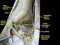
 Coronal section through right ankle and subtalar joints. (Label for Peroneus brevis is at right, third from the bottom.)
 -
 Bones of the right leg, anterior surface.
 -
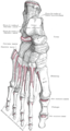 Bones of the right foot, dorsal surface.
 -
 Muscles of the front of the leg.
 -
 Cross-section through middle of leg.
 -
 The popliteal, posterior tibial, and fibular arteries.
 -
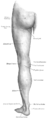 Back of left lower extremity.
 -
 Fibularis brevis muscle
 -
 Muscles of the sole of the foot.
 -
 Dorsum of foot, deep dissection.
 -
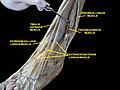 Muscles of leg, lateral view, deep dissection.


See also
References
![]() This article incorporates text in the public domain from page 487 of the 20th edition of Gray's Anatomy (1918)
This article incorporates text in the public domain from page 487 of the 20th edition of Gray's Anatomy (1918)
- ISBN 978-0-12-410390-0, retrieved 2021-02-20
- ISBN 978-0-323-02358-0, retrieved 2021-02-20
- ^ a b c Gray's Anatomy (1918), see infobox
- ^ FIPAT (2019). Terminologia Anatomica. Federative International Programme for Anatomical Terminology.
External links
- Anatomy photo:15:st-0407 at the SUNY Downstate Medical Center
- PTCentral