
Popliteus muscle
This article includes a list of general references, but it lacks sufficient corresponding inline citations. (May 2015) |
| Popliteus muscle | |
|---|---|
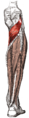
 Popliteus muscle (shown in red) | |
Dissection video (2 min 22 s) | |
| Details | |
| Origin | lateral femoral epicondyle |
| Insertion | posterior surface of the tibia proximal to the soleus line |
| Artery | Popliteal artery |
| Nerve | Tibial nerve |
| Actions | Medially rotates tibia on the femur if the femur is fixed (sitting down) or laterally rotates femur on the tibia if tibia is fixed (standing up), unlocks the knee to allow flexion (bending), helps to prevent the forward dislocation of the femur while crouching |
| Identifiers | |
| Latin | musculus popliteus, poplit=ham (pit) of the knee |
| TA98 | A04.7.02.050 |
| TA2 | 2665 |
| FMA | 22590 |
| Anatomical terms of muscle | |
The popliteus muscle in the leg is used for unlocking the
Structure
The popliteus muscle originates from the lateral surface of the
Nerve supply
The popliteus muscle is supplied by the
Variation
There is sometimes an additional head from the sesamoid bone in the lateral (outer) head of the gastrocnemius muscle.
Rarely an additional inconstant muscle; the popliteus minor is seen. It originates from the femur on the inner side of the plantaris muscle and inserts into the posterior ligament of the knee-joint.
Peroneotibialis, 14% of population. Origin is inner side of the head of the fibula, insertion into the upper end of the oblique line of the tibia, it lies beneath the popliteus.[3]
Another variant, the cyamella, is a small sesamoid bone embedded in the tendon of the popliteus muscle. It is rarely seen in humans, with prevalence rates from 0.57–1.8%,[4] but has been described more often in other primates and certain other animals.[5]
Function
The popliteus assists in flexing the leg upon the thigh; when the leg is flexed, it will rotate the tibia inward.
It is especially called into action at the beginning of the act of bending the knee, in as much as it produces the slight
When the knee is in full extension, the femur slightly medially rotates on the tibia to lock the knee joint in place. Popliteus is often referred to as the "Key" to unlocking the knee since it begins knee flexion by laterally rotating the femur on the tibia.[6]
Popliteus is also attached to the lateral meniscus in the knee and draws it posteriorly during knee flexion to prevent crushing the meniscus between the tibia and femur as the knee flexes.
Additional images
-
 Animation
 -
 Deep layer of muscles on the back of the right leg
 -
 Muscles ofdeep posterior compartmentof the right leg

See also
- Posterolateral knee
Surgery
Injury to the Popliteus causes posterolateral rotatory instability of knee. This can be treated with Arthroscopic Popliteus Sling reconstruction using the popliteus portal.[7]
References
![]() This article incorporates text in the public domain from page 484 of the 20th edition of Gray's Anatomy (1918)
This article incorporates text in the public domain from page 484 of the 20th edition of Gray's Anatomy (1918)
- ^ ISBN 978-0-323-32903-3, retrieved 2021-03-01
- PMID 24617694.
- ^ Gray, Henry. 1918. Anatomy of the Human Body. Page 485
- S2CID 233427472.
- S2CID 13339926.
- ^ S2CID 210831808.
- S2CID 76366926.
External links
- Anatomy photo:15:st-0413 at the SUNY Downstate Medical Center
- PTCentral