
Gluteus maximus
| Gluteus maximus | |
|---|---|
psoas minor | |
| Identifiers | |
| Latin | musculus glutaeus maximus |
| TA98 | A04.7.02.006 |
| TA2 | 2598 |
| FMA | 22314 |
| Anatomical terms of muscle] | |
The gluteus maximus is the main
Its large size is one of the most characteristic features of the muscular system in humans,[2] connected as it is with the power of maintaining the trunk in the erect posture. Other primates have much flatter hips and cannot sustain standing erectly.
The muscle is made up of muscle fascicles lying parallel with one another, and are collected together into larger bundles separated by fibrous septa.
Structure

The gluteus maximus (or buttock) is the outermost muscle of the buttocks. It arises from connections to nearby structures in this area. It arises from the posterior gluteal line of the inner upper
The fibers are directed obliquely inferiorly and laterally;
The gluteus maximus ends in two main areas:
- those forming the upper and larger portion of the muscle, together with the superficial fibers of the lower portion, end in a thick tendinous lamina, which passes across the greater trochanter, and inserts into the iliotibial band of the fascia lata;
- the deeper fibers of the lower portion are inserted into the adductor magnus. If present, the third trochanteralso serves as an attachment.
Bursae
Three
- One of these, of large size, separates it from the greater trochanter;
- a second (often missing) is situated on the tuberosity of the ischium;
- a third is found between the tendon of the muscle and that of the vastus lateralis.
-
 Image showing the outer surface of the ilium, showing the inferior gluteal line.

Function
The gluteus maximus straightens the leg at the hip; when the leg is flexed at the hip, the gluteus maximus extends it to bring the leg into a straight line with the body.[3] The anus also aligns when the leg is flexed at the hip, making the muscle tighten and the pelvis tilt forward.
Taking its fixed point from below, it acts upon the
The gluteus maximus is a tensor of the
The lower part of the muscle also acts as an
Society and culture
Training


The gluteus maximus is involved in several sports, from running to weight-lifting. A number of exercises focus on the gluteus maximus and other muscles of the upper leg:
- Hip thrusts
- Glute bridge
- Quadruped hip extensions
- Kettlebell swings
- Squats and variations like split squats, unilateral squats with the raised foot positioned either backwards or forwards (pistols), and wide-stance lunges
- Deadlift (and variations)
- Reverse hyperextension
- Four-way hip extensions
- Glute-ham raise
In art
In cultural terms, the glutes are often considered a symbol of health and strength, and aesthetically appealing. They frequently feature in artwork which seeks to emphasise and celebrate physicality, and the ability to move dynamically and powerfully. They are usually shown to be efficiently proportioned and prominent.
Evidence of such depictions of the gluteal muscles extends from at least Ancient Greece to the modern day.[5][6]
- The glutes in art
-
 An Ancient Greek javelin thrower represented on a vase, c.520 B.C.
 -
 An Ancient Greek warrior in bronze.Riace Bronzes, c.450 B.C.


Clinical significance
Functional assessment can be useful in assessing injuries to the gluteus maximus and surrounding muscles.
The 30-second chair-to-stand test measures a participant's ability to stand up from a seated position as many times as possible in a thirty-second period of time.[7] Testing the number of times a person can stand up in a thirty-second period helps assess strength, flexibility, pain, and endurance,[7] which can help determine how far along a person is in rehabilitation, or how much work is still to be done.
The
Other animals
The gluteus maximus is larger in size and thicker in humans than in other primates.[3] Its large size is one of the most characteristic features of the muscular system in humans,[2] connected as it is with the power of maintaining the trunk in the erect posture. Other primates have much flatter hips and cannot sustain standing erectly.[9][10]
In other primates, the correlate to the human gluteus maximus consists of the ischiofemoralis, a small muscle that corresponds to the human gluteus maximus and originates from the ilium and the ligaments of the sacroiliac, and the gluteus maximus proprius, a large muscle that extends from the ischial tuberosity to a relatively more distant insertion on the femur. In adapting to bipedal gait, reorganization of the attachment of the muscle as well as the moment arm was required.[9]
Additional images
-
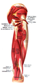
 Gluteus maximus is the most superficial muscle of thehips, here visible at top centre with skin removed from the entire right leg
 -
 All gluteal muscles, maximus in yellow
 -
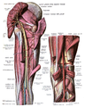
 Structures surrounding righthip-joint(gluteus maximus visible at bottom)
 -
 Innervation and blood-supply of the gluteus maximus.
 -
 Gluteus maximus cut showing underlying structures.
 -
 Structures visible under the gluteus maximus.
 -
 Innervation as seen from under the gluteus maximus.
 -
 The gluteus maximus, with surrounding fascia. Right buttock, viewed from behind, skin covering removed.


See also
References
![]() This article incorporates text in the public domain from page 474 of the 20th edition of Gray's Anatomy (1918)
This article incorporates text in the public domain from page 474 of the 20th edition of Gray's Anatomy (1918)
- ^ "What is the strongest muscle in the human body?". Library of Congress. 19 November 2019. Retrieved 2023-05-28.
- ^ a b Norman Eizenberg et al., General Anatomy: Principles and Applications (2008), p. 17.
- ^ OCLC 920806541.
- ISBN 978-1628603-46-0.
- ISBN 9781628603460.
- ISBN 0773512314.
- ^ a b
Dobson, F.; Bennell, K.; Hinman, R.; Abbott, H.; Roos, E. (2013). "OARSI recommended performance-based tests to assess physical function in people diagnosed with hip or knee osteoarthritis" (PDF). Osteoarthritis and Cartilage. 21 (8): 1042–52. PMID 23680877.
- ^ "Passive Piriformis ROM". Exrx.net. Retrieved February 19, 2015.
- ^ PMID 27011809.
- .
External links
- Cross section image: pelvis/pelvis-female-17—Plastination Laboratory at the Medical University of Vienna
- Cross section image: pelvis/pelvis-e12-15—Plastination Laboratory at the Medical University of Vienna
- Cross section image: pembody/body18b—Plastination Laboratory at the Medical University of Vienna