
Gluteus minimus
| Gluteus minimus | ||
|---|---|---|
Antagonist Lateral rotator group | | |
| Identifiers | ||
| Latin | musculus glutaeus minimus | |
| TA98 | A04.7.02.008 | |
| TA2 | 2600 | |
| FMA | 22317 | |
| Anatomical terms of muscle] | ||
The gluteus minimus, or glutæus minimus, the smallest of the three
Structure

It is fan-shaped, arising from the outer surface of the ilium, between the anterior and inferior gluteal lines, and behind, from the margin of the greater sciatic notch.
The fibers converge to the deep surface of a radiated
Relations
A
Between the
The deep surface of the gluteus minimus is in relation with the reflected tendon of the rectus femoris and the capsule of the hip joint.
Variations
The muscle may be divided into an anterior and a posterior part, or it may send slips to the
Function
The gluteus medius and gluteus minimus abduct the thigh, when the limb is extended, and are principally called into action in supporting the body on one limb, in conjunction with the tensor fasciæ latæ.
Their anterior fibers also flex the hip, and by drawing the greater trochanter forward, rotate the thigh inward,[2][3] in which action they are also assisted by the Tensor fasciæ latæ.
Both gluteus minimus and medius have the same function. Their primary function is abduction of the femur, while internal rotation and flexion can occur depending on the position of the femur.[4] Additionally, with the hip flexed, the gluteus minimus internally rotates the thigh. With the hip extended, gluteus minimus externally rotates the thigh.[4][5]
The attachment to the superior capsule of the hip may also serve to retract the capsule away from the joint during motion. This mechanism may prevent capsular impingement similar to the role of the articularis genus in the knee.[6]
Clinical significance
Paralysis of this muscle or gluteus medius, such as may be caused by the superior gluteal nerve palsy, can lead to difficulty abducting the leg. People will compensate for their difficulty walking by adopting a Trendelenburg gait.
References
![]() This article incorporates text in the public domain from page 475 of the 20th edition of Gray's Anatomy (1918)
This article incorporates text in the public domain from page 475 of the 20th edition of Gray's Anatomy (1918)
- )
- S2CID 1236412.
- ISBN 9780434902156.
- ^ .
- ^ Pratt, N. Clinical Musculoskeletal Anatomy. CBLS: Marietta, OH 2004.
- ^ Neuman, Donald. Kinesiology of the Musculoskeletal System. pp. 494–495.
Additional images
-
 Position of gluteus minimus muscle (shown in red). Hip bone is shown in semi-transparent.
 -
 Structures surrounding right hip-joint. (Gluteus minimus visible at center left.)
 -
 Right hip bone. External surface.
 -
 Right femur. Anterior surface.
 -
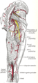 The arteries of the gluteal and posterior femoral regions.
 -
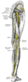 Nerves of the right lower extremity Posterior view.


External links
- PTCentral
- Anatomy photo:13:st-0406 at the SUNY Downstate Medical Center