
Pectineus muscle
| Pectineus | |
|---|---|
adduction, external rotation | |
| Identifiers | |
| Latin | musculus pectineus |
| TA98 | A04.7.02.025 |
| TA2 | 2627 |
| FMA | 22440 |
| Anatomical terms of muscle] | |
The pectineus muscle (
It can be classified in the medial compartment of thigh[2] (when the function is emphasized) or the anterior compartment of thigh (when the nerve is emphasized).[3]
Structure
The pectineus muscle arises from the
Relations
The pectineus is in relation by its anterior surface with the pubic portion of the
By its posterior surface with the
By its external border with the
By its internal border with the outer edge of the
Obturator foramen is situated directly behind this muscle, which forms one of its coverings.[4]
It forms part of the floor of the femoral triangle.
Innervation
The lumbar plexus is formed from the anterior rami of nerves L1 to L4 and some fibers from T12. With only five roots and two divisions, it is less complex than the brachial plexus and gives rise to a number of nerves including the femoral nerve and accessory obturator nerve. The pectineus muscle is considered a composite muscle as the innervation is by the femoral nerve (L2 and L3) and occasionally (20% of the population) a branch of the obturator nerve called the accessory obturator nerve. When it is present, the accessory obturator nerve innervates a portion of the pectineus muscle, entering the muscle on its dorsomedial aspect. The greater nerve to the muscle is the femoral nerve. Unlike the obturator accessory nerve, the femoral nerve is always present and provides the sole innervation for the pectineus muscle in over 90% of cases. The muscle is also innervated by the accessory obturator nerve in the 8.7% of cases in which the nerve occurs.[5]
Function
Its primary functions are contributing to
Additional images
-
 Right hip bone. External surface.
 -
 Structures surrounding right hip-joint.
 -
 Muscles of the iliac and anterior femoral regions.
 -
 Deep muscles of the medial femoral region.
 -
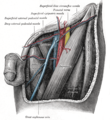 The left femoral triangle.
 -
 The lumbar plexus and its branches.
 -
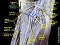
 Pectineus muscle
 -
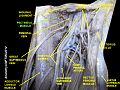
 Pectineus muscle
 -
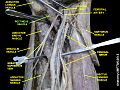
 Pectineus muscle
 -
 Pectineus muscle
 -
 Pectineus muscle
 -
 Muscles of Thigh. Anterior views
 -
 Muscles of Thigh. Anterior views.


See also
References
![]() This article incorporates text in the public domain from page 472 of the 20th edition of Gray's Anatomy (1918)
This article incorporates text in the public domain from page 472 of the 20th edition of Gray's Anatomy (1918)
- Mosby's Medical, Nursing & Allied Health Dictionary, Fourth Edition, Mosby-Year Book Inc., 1994, p. 1177
- ISBN 0-443-07168-3.
- ^ medialthigh at The Anatomy Lesson by Wesley Norman (Georgetown University)
- ^ Wilson, Erasmus (1851). The anatomist's vade mecum: a system of human anatomy. John Churchill. p. 260.
- ^ RUSSELL T., WOODBURNE. "The Accessory Obturator Nerve and the Innervation of the Pectineus Muscle" (PDF): 367.
{{cite journal}}: Cite journal requires|journal=(help) - PMID 20118525.
Notes
- Woodburne, Russell (1960). "The Accessory Obturator Nerve and the Innervation of the Pectineus Muscle" (PDF). Michigan Library Med School. 136 (3): 367–369. S2CID 14846721. Retrieved 2 December 2015.
- Saladin, Kenneth S. Anatomy & Physiology: The Unity of Form and Function. New York, NY: McGraw-Hill, 2007. pg.493. Print.
External links
- Anatomy figure: 12:02-05 at Human Anatomy Online, SUNY Downstate Medical Center - "Muscles of the anterior (extensor) compartment of the thigh."
- Anatomy figure: 12:03-04 at Human Anatomy Online, SUNY Downstate Medical Center - "Deep muscles of the anterior thigh."
- Cross section image: pelvis/pelvis-e12-15—Plastination Laboratory at the Medical University of Vienna