
Biceps femoris muscle
| Biceps femoris | |
|---|---|
Quadriceps muscle | |
| Identifiers | |
| Latin | musculus biceps femoris |
| TA98 | A04.7.02.032 |
| TA2 | 2638 |
| FMA | 22356 |
| Anatomical terms of muscle] | |
The biceps femoris (
Structure
It has two heads of origin:
- the long head arises from the lower and inner impression on the posterior part of the tuberosity of the ischium. This is a common tendon origin with the semitendinosus muscle, and from the lower part of the sacrotuberous ligament.[2]
- the short head, arises from the lateral lip of the gluteus maximus, from the lateral prolongation of the linea aspera to within 5 cm. of the lateral condyle; and from the lateral intermuscular septum.[2]
The two muscle heads joint together distally and unite in an intricate fashion. The fibers of the long head form a fusiform belly, which passes obliquely downward and lateralward across the sciatic nerve to end in an aponeurosis which covers the posterior surface of the muscle and receives the fibers of the short head. Inferiorly, the aponeurosis condenses to form a tendon which predominantly inserts onto the lateral side of the head of the fibula. There is a second small insertional attachment by a small tendon slip into the lateral condyle of the tibia.[2]
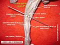
At its insertion the tendon divides into two portions, which embrace the fibular collateral ligament of the knee-joint. Together, this joining of tendons is commonly referred to as the conjoined tendon of the knee.[2][3]
From the posterior border of the tendon a thin expansion is given off to the fascia of the leg. The tendon of insertion of this muscle forms the lateral hamstring; the common fibular (peroneal) nerve descends along its medial border.[2]
Variations
The short head may be absent; additional heads may arise from the
A slip may pass to the gastrocnemius.[2]
Innervation
It is a composite muscle as the short head of the biceps femoris develops in the flexor compartment of the thigh and is thus innervated by common fibular branch of the sciatic nerve (L5, S1), while the long head is innervated by the tibial branch of the sciatic nerve (L5, S1).[5]
Blood supply
The muscle's vascular supply is derived from the
Function
Both heads of the biceps femoris perform knee flexion.[6]
Since the long head originates in the pelvis it is involved in hip extension.
When the knee is semi-flexed, the biceps femoris in consequence of its oblique direction rotates the leg slightly outward.
Clinical significance
See also
Additional images
-
 Right hip bone. External surface.
 -
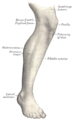
 Bones of the right leg. Anterior surface.
 -
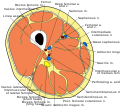 Cross-section through the middle of the thigh.
 -
 Muscles of the gluteal and posterior femoral regions.
 -
 The popliteal, posterior tibial, and peroneal arteries.
 -
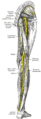 Nerves of the right lower extremity Posterior view.
 -
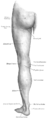 Back of left lower extremity.
 -
 Biceps femoris


References
![]() This article incorporates text in the public domain from page 478 of the 20th edition of Gray's Anatomy (1918)
This article incorporates text in the public domain from page 478 of the 20th edition of Gray's Anatomy (1918)
- ^ Rodgers, Cooper D.; Raja, Avais. StatPearls - Anatomy, Bony Pelvis and Lower Limb, Hamstring Muscle. StatPearls Publishing. Archived from the original on 25 February 2023. Retrieved 5 April 2023.
- ^ a b c d e f g "Gray's Anatomy". 1918. Archived from the original on 2009-12-22. Retrieved 2010-09-01.
- ^ Smithius, Robin; Rubin, David (August 2, 2005). "Knee - Non-Meniscal pathology". The Radiology Assistant.
- ^ The Adult Knee, vol. 1, ed. Callaghan, p. 70
- ^ a b "biceps femoris muscle (anatomy)". GPnotebook.
- ^ a b Origin, insertion and nerve supply of the muscle at Loyola University Chicago Stritch School of Medicine
Further reading
- Kumakura, Hiroo (July 1989). "Functional analysis of the biceps femoris muscle during locomotor behavior in some primates". American Journal of Physical Anthropology. 79 (3): 379–391. PMID 2504047.
- Marshall, John L.; Girgis, Fakhry G.; Zelko, Russel R. (1972). "The Biceps Femoris Tendon and Its Functional Significance". J Bone Joint Surg Am. 54 (7): 1444–1450. PMID 4653628. Archived from the originalon 2008-10-10. Retrieved 2010-09-01.
- Sneath, R. S. (October 1955). "The insertion of the biceps femoris". J. Anat. 89 (89(Pt 4)): 550–553. PMID 13278305.
External links
- UWash - long head
- UWash - short head
- Anatomy photo:14:06-0100 at the SUNY Downstate Medical Center
- Anatomy photo:14:st-0402 at the SUNY Downstate Medical Center