
Preoptic area
| Preoptic area | |
|---|---|
 Preoptic area is 'PO', at left, in blue. | |
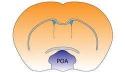 Preoptic area of the mouse brain | |
| Details | |
| Identifiers | |
| Latin | area praeoptica |
| Acronym(s) | POA |
| MeSH | D011301 |
| NeuroNames | 377 |
| NeuroLex ID | birnlex_1706 |
| TA98 | A14.1.08.407 |
| TA2 | 5710 |
| FMA | 62313 |
| Anatomical terms of neuroanatomy | |
The preoptic area is a region of the hypothalamus. MeSH classifies it as part of the anterior hypothalamus. TA lists four nuclei in this region, (medial, median, lateral, and periventricular).
Functions
The preoptic area is responsible for
Nuclei
Median preoptic nucleus
The median preoptic nucleus is located along the midline in a position significantly dorsal to the other three preoptic nuclei, at least in the crab-eating macaque brain. It wraps around the top (dorsal), front, and bottom (ventral) surfaces of the anterior commissure.
The median preoptic nucleus generates
Medial preoptic nucleus
The medial preoptic nucleus is bounded laterally by the lateral preoptic nucleus, and medially by the preoptic periventricular nucleus. It releases gonadotropin-releasing hormone (GnRH), controls copulation in males, and is larger in males than in females.
Parental behavior
The medial preoptic area (mPOA) has been implicated in parental care in both males and females. In rats,
Sexual behavior
The mPOA is sexually dimorphic, that is, it differs in function between males and females. In females, studies have examined the influence of the mPOA on precopulatory and appetitive behaviors. Precopulatory behaviors involve several brain areas, including the mPOA as well as the medial
In male rats, the mPOA affects the consummatory phase of sexual behavior, and possibly motivation, with lesions causing a complete loss of copulatory behaviors.[9] Conversely, electrical stimulation of this area triggers male copulatory behavior, as measured by decreases in the latency to ejaculate.[10] Furthermore, testosterone implanted into the mPOA of castrated males completely restores mating, as long as aromatase is not inhibited.[11]
Ventrolateral preoptic nucleus
The ventrolateral preoptic nucleus, or intermediate nucleus, is adjacent to the medial preoptic nucleus. It also mediates non-REM sleep onset.
Preoptic periventricular nucleus
The preoptic periventricular nucleus is located along the midline and is medial to the medial preoptic nucleus.