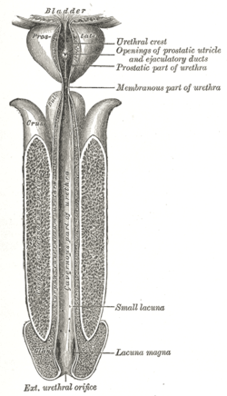
Seminal colliculus
| Seminal colliculus | |
|---|---|
ductus deferentes, seen from the front. | |
 The male urethra laid open on its anterior (upper) surface. | |
| Details | |
| Identifiers | |
| Latin | colliculus seminalis, verumontanum |
| TA98 | A09.4.02.008 |
| TA2 | 3448 |
| FMA | 74363 |
| Anatomical terminology] | |
The seminal colliculus (
urothelium
that characterizes the landmark on magnified views.
Embryologically, it is derived from the uterovaginal primordium. The landmark is important in classification of several urethral developmental disorders. The margins of seminal colliculus are the following:
- the orifices of the prostatic utricle
- the slit-like openings of the ejaculatory ducts.
- the openings of the prostatic ducts

Posterior urethral valves
The verumontanum is an important anatomic landmark for pathology in a
hypospadia disorders and is then seen in the bulbous, or penile portion of the urethra.[3]
Prostatic utricle
The prostatic utricle (embryologic derivative of urogenital sinus and the male vestigial equivalent of vagina) arises from the urethra at the level of the verumontanum and projects posteriorly. This blind ending structure can be associated with hypospadias. This is distinct from a Cowper duct syringocele, which arises at the bulbous urethra.
References
- ^ "Posterior Urethral Valve. Emedicine, Radiology". eMedicine. July 18, 2010. Retrieved July 18, 2010.
- ^ Maxwell Smith, M Scott Lucia, Priya N Werahera and Francisco G La Rosa Carcinoid tumor of the verumontanum (colliculus seminalis) of the prostatic urethra with a coexisting prostatic adenocarcinoma: a case report Journal of Medical Case Reports 2010, 4:16
- ^ F Ikoma, H Shima. 1991. Caudal migration of the verumontanum. Journal of Pediatric Surgery. 26: 7, 858-861.
External links
- Anatomy photo:44:05-0202 at the SUNY Downstate Medical Center — "The Male Pelvis: The Prostate Gland"
- pelvis at The Anatomy Lesson by Wesley Norman (Georgetown University) (malebladder)
- figures/chapter_34/34-3.HTM: Basic Human Anatomy at Dartmouth Medical School
