
Seminiferous tubule
| Seminiferous tubule | |
|---|---|
Efferent ductules 6: Rete testis | |
| Details | |
| Identifiers | |
| Latin | tubuli seminiferi |
| MeSH | D012671 |
| TA98 | A09.3.01.022 |
| TA2 | 3599 |
| FMA | 19825 |
| Anatomical terminology] | |
Seminiferous tubules are located within the
spermatozoa
.
Structure
The epithelium of the tubule consists of a type of sustentacular cells known as Sertoli cells, which are tall, columnar type cells that line the tubule.
In between the Sertoli cells are
androgen-binding protein
, a binding protein which increases the concentration of testosterone.
There are two types: convoluted and straight, convoluted toward the lateral side, and straight as the tubule comes medially to form ducts that will exit the testis.
The seminiferous tubules are formed from the testis cords that develop from the primitive
gonadal ridge
.
Function
enzymes employed in these repair processes may lead to infertility.[2]
Additional images
-
-
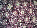
 Longitudinal section through the left side of the scrotum and the left testis (seminiferous tubules visible in center, but not labeled).
 -
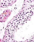
 Seminiferous tubule (transverse section).
 -
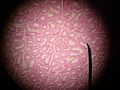
 Photomicrograph of section through rat testis, showing seminiferous tubules.


See also
References
External links
- Histology image: 17802loa – Histology Learning System at Boston University
- Image
- Diagram Archived 2007-02-19 at the Wayback Machine