
Rete testis
| Rete testis | |
|---|---|
Wolffian duct | |
| Identifiers | |
| Latin | rete testis |
| MeSH | D012152 |
| TA98 | A09.3.01.024 |
| TA2 | 3601 |
| FMA | 19834 |
| Anatomical terminology] | |
The rete testis ( Its function is to provide a site for fluid reabsorption.
Structure

The rete testis is the network of interconnecting tubules where the
Development
In the
Function
It appears the function of the rete testis is to mix the sperm as they leave the seminiferous tubules. Sperm leave the seminiferous tubules in the dilute secretions of Sertoli cells. The rete testis does modify the luminal fluids with a limited amount of secretion and reabsorption, but their primary function is to mix and transport the sperm into the efferent ductules, where the major function is reabsorption of about 95% of the fluid, which increases the sperm concentration prior to entering the epididymis.
Clinical significance
Etymology
English uses the Neo-Latin name for the structure, which simply means "network of the testis".
Additional images
-
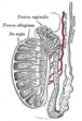 Vertical section of thetestis, to show the arrangement of the ducts.
 -
 Micrograph of the rete testis involved by seminoma. H&E stain.
 -
 Tubular ectasia of the rete testis


References
External links
- Image at UC Davis Archived 2006-04-09 at the Wayback Machine
- Diagram Archived 2016-03-04 at the Wayback Machine
- Diagram
This article related to the genitourinary system is a stub. You can help Wikipedia by expanding it. |