
Postcentral gyrus
| Postcentral gyrus | |
|---|---|
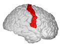
 Postcentral gyrus of the human brain | |
 Brodmann areas 3, 1 and 2 of human brain. Brodmann area 3 is in red, area 1 in green, and area 2 in yellow. | |
| Details | |
| System | Somatosensory system |
| Location | Parietal lobe |
| Artery | Middle cerebral artery |
| Function | Primary somatosensory cortex |
| Identifiers | |
| Latin | gyrus postcentralis |
| NeuroNames | 105 |
| NeuroLex ID | birnlex_1070 |
| TA98 | A14.1.09.128 |
| TA2 | 5469 |
| FMA | 61896 |
| Anatomical terms of neuroanatomy | |
In
sensory homunculus
.
The primary somatosensory cortex was initially defined from surface stimulation studies of
2, more recent work by Kaas has suggested that for homogeny with other sensory fields only area 3 should be referred to as "primary somatosensory cortex", as it receives the bulk of the thalamocortical projections from the sensory input fields[citation needed
].
Structure
The lateral postcentral gyrus is bounded by:
- medial longitudinal fissure medially(to the middle)
- central sulcus rostrally (in front)
- postcentral sulcus caudally (in back)
- lateral sulcus inferiorly (underneath)
The postcentral gyrus includes Brodmann areas 1, 2, and 3. Brodmann area 1 occupies the apex of the postcentral gyrus.
See also
Additional images
-
 Postcentral gyrus (animation)
 -
 Lateral surface of left cerebral hemisphere, viewed from the side.
 -
 Primary cortices, including primary somatosensory cortex (labeled in purple)
 -
 Postcentral gyrus, showed on the right hemisphere.
 -
 Postcentral gyrus highlighted in green on coronal T1 MRI images
 -
 Postcentral gyrus highlighted in green on sagittal T1 MRI images
 -
 Postcentral gyrus highlighted in green on transversal T1 MRI images


External links
Wikimedia Commons has media related to Postcentral gyrus.
- ancil-1040 at NeuroNames - area 1
- ancil-1041 at NeuroNames - area 2
- ancil-1042 at NeuroNames - area 3