
Cerebral hemisphere
| Cerebral hemisphere | |
|---|---|
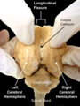
 Human brain seen from front. | |
 Right cerebral hemisphere Left cerebral hemisphere | |
| Details | |
| Identifiers | |
| Latin | hemisphaerium cerebri |
| NeuroNames | 241 |
| NeuroLex ID | birnlex_1796 |
| TA98 | A14.1.09.002 |
| TA2 | 5418 |
| FMA | 61817 |
| Anatomical terms of neuroanatomy | |
The
There are three known poles of the cerebral hemispheres: the occipital pole, the frontal pole, and the temporal pole.
The central sulcus is a prominent fissure which separates the parietal lobe from the frontal lobe and the primary motor cortex from the primary somatosensory cortex.
Structure
Each cerebral hemisphere has an outer layer of
Poles
_-_inferiror_view.png)
There are three poles of the cerebrum: the occipital pole, the frontal pole, and the temporal pole. The occipital pole is the posterior end of each occipital lobe in each hemisphere. It is more pointed than the rounder frontal pole. The frontal pole is at the frontmost part of the frontal lobe in each hemisphere, and is more rounded than the occipital pole. The temporal pole is located between the frontal and occipital poles, and sits in the anterior part of middle cranial fossa in each temporal lobe.[5]
Composition
If the upper part of either hemisphere is removed, at a level about 1.25 cm above the corpus callosum, the central white matter will be exposed as an oval-shaped area, the centrum semiovale, surrounded by a narrow convoluted margin of gray substance, and studded with numerous minute red dots (puncta vasculosa), produced by the escape of blood from divided blood vessels.[citation needed]
If the remaining portions of the hemispheres be slightly drawn apart a broad band of white substance, the corpus callosum, will be observed, connecting them at the bottom of the longitudinal fissure; the margins of the hemispheres which overlap the corpus callosum are called the labia cerebri.[6]
Each labium is part of the cingulate gyrus already described; and the groove between it and the upper surface of the corpus callosum is termed the
If the hemispheres are sliced off to a level with the upper surface of the corpus callosum, the white substance of that structure will be seen connecting the two hemispheres.
The large expanse of medullary matter now exposed, surrounded by the convoluted margin of gray substance, is called the centrum semiovale. The blood supply to the centrum semiovale is from the superficial middle cerebral artery.[3] The cortical branches of this artery descend to provide blood to the centrum semiovale.[7]
Development
The cerebral hemispheres are derived from the
Function
Hemisphere lateralization
Broad generalizations are often made in popular psychology about certain functions (e.g. logic, creativity) being lateralized, that is, located in the right or left side of the brain. These claims are often inaccurate, as most brain functions are actually distributed across both hemispheres. Most scientific evidence for asymmetry relates to low-level perceptual functions rather than the higher-level functions popularly discussed (e.g. subconscious processing of grammar, not "logical thinking" in general).[8][9] In addition to this lateralization of some functions, the low-level representations also tend to represent the contralateral side of the body.
The best example of an established lateralization is that of Broca's and Wernicke's Areas (

Perceptual information is processed in both hemispheres, but is laterally partitioned: information from each side of the body is sent to the opposite hemisphere (visual information is partitioned somewhat differently, but still lateralized). Similarly, motor control signals sent out to the body also come from the hemisphere on the opposite side. Thus, hand preference (which hand someone prefers to use) is also related to hemisphere lateralization.[citation needed]
In some aspects, the hemispheres are asymmetrical; the right side is slightly bigger. There are higher levels of the neurotransmitter norepinephrine on the right and higher levels of dopamine on the left. The right hemisphere is more sensitive to testosterone. There is more white matter (longer axons) on the right and more grey matter (cell bodies) on the left.[12]
Clinical significance
Infarcts of the centrum ovale can occur.[3]
As a treatment for epilepsy the corpus callosum may be severed to cut the major connection between the hemispheres in a procedure known as a corpus callosotomy.
A
Additional images
-
 The sheep brain seen from the back. Opening longitudinal fissure which separates left and right cerebral hemispheres.
 -
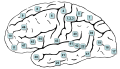 Lateral surface. (The frontal pole is approximately at 10, the occipital pole is approximately at 17, and the temporal pole is approximately at 38.)


References
- PMID 9010002.
- S2CID 15968665.
- ^ S2CID 219195107.
- ISBN 978-0-7817-9427-5.
- ISBN 978-81-312-3727-4.
- ISBN 9781317705703. Retrieved 13 August 2019.
- PMID 16291890.
- ISBN 978-0-470-80552-7.
- ^ "Neuromyth 6: The left brain/ right brain myth". Organisation for Economic Co-operation and Development (OECD). Retrieved 15 October 2011.
- S2CID 16060074.
- PMID 26766393.
- ISBN 978-0-520-22461-2.
- PMID 10320379.
| National | |
|---|---|
| Other | |