
Bile duct hamartoma
| Bile duct hamartoma | |
|---|---|
 | |
| Histopathology of a bile duct hamartoma, low magnification, H&E stain, showing a well demarcated lesion. |
Bile duct hamartoma or biliary hamartoma, are benign lesions of the intrahepatic bile duct.[1] They are classically associated with polycystic liver disease, as may be seen in the context of polycystic kidney disease, and represent a malformation of the liver plate.[2]
Signs and symptoms
Most patients are asymptomatic. When patients do present with symptoms the most common symptom is abdominal pain. Other symptoms include fever, weight loss, and jaundice.[1]
Causes
Biliary duct hamartomas are defects resulting from the failure of embryonic bile duct involution that affect the small interlobular bile ducts. Patients with polycystic kidney disease and polycystic liver are far more likely to have them.[1]
Diagnosis
-
 Histopathology of a bile duct hamartoma, high magnification, H&E stain. It shows typical features of bile duct hamartoma: Small to medium sized, irregularly shaped bile ducts lined by bland cuboidal epithelium (may also be flattened). Prominent intervening collagenous stroma. Bile ducts containing eosinophilic debris (may also contain inspissated bile)
 -
 von Meyenburg Complex in ultrasound. Numerous little cysts with ringdown artefacts.


Laboratory findings include high transaminase levels, raised gamma-glutamyl transferase or alkaline phosphatase levels, increased C-reactive protein, hypoalbuminemia, and hematologic abnormalities like thrombocytopenia, leukopenia, leukocytosis, and anemia.[1]
At
Eponym
The eponymous terms (
Additional images
-
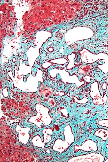 Trichrome stain. Intermediate magnification
 -
 Trichrome stain, high magnification
 -
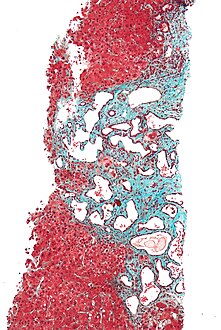 Low magnificationTrichrome stain.
 -
 Gross pathologic appearance of a large bile duct hamartoma.


See also
References
Further reading
- Zheng, Rong Qin; Kudo, Masatoshi; Onda, Hirokazu; Inoue, Tatsuo; Maekawa, Kiyoshi; Minami, Yasunori; Chung, Hobyung; Kitano, Masayuki; Kawasaki, Toshihiko (2005-12-19). "Imaging findings of biliary hamartomas (von Meyenburg complexes)". Journal of Medical Ultrasonics. 32 (4): 205–212. S2CID 25856248.