
Middle frontal gyrus
| Middle frontal gyrus | |
|---|---|
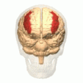
 Middle frontal gyrus of the human brain. | |
 Coronal section through anterior cornua of lateral ventricles. | |
| Details | |
| Part of | Frontal lobe |
| Artery | Middle cerebral |
| Identifiers | |
| Latin | gyrus frontalis medius |
| NeuroNames | 84 |
| NeuroLex ID | birnlex_1451 |
| TA98 | A14.1.09.118 |
| TA2 | 5454 |
| FMA | 61859 |
| Anatomical terms of neuroanatomy | |
The middle frontal gyrus makes up about one-third of the frontal lobe of the human brain. (A gyrus is one of the prominent "bumps" or "ridges" on the surface of the human brain.)
The middle frontal gyrus, like the
frontal gyrus
than a true gyrus.
The borders of the middle frontal gyrus are the inferior frontal sulcus below; the superior frontal sulcus above; and the precentral sulcus behind.[1]
Additional images
-
 Position of middle frontal gyrus (shown in red).
 -
 Left cerebral hemisphere seen from above.
 -
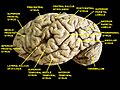
 Lateral surface of left cerebral hemisphere.
 -
 Lateral surface of right cerebral hemisphere. Middle frontal gyrus is noted by red arrows.
 -
 Cerebrum. Lateral view.Deep dissection.
 -
 Cerebrum. Lateral view.Deep dissection.
 -
 Cerebrum. Lateral view.Deep dissection.
 -
 Rostral middle frontal gyrus.
 -
 Caudal middle frontal gyrus.
 -
 Middle frontal gyrus highlighted in green on coronal T1 MRI images
 -
 Middle frontal gyrus highlighted in green on sagittal T1 MRI images
 -
 Middle frontal gyrus highlighted in green on transversal T1 MRI images


References
- S2CID 232310374.
Wikimedia Commons has media related to Middle frontal gyrus.