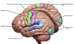
Primary somatosensory cortex
| Primary somatosensory cortex | |
|---|---|
 Primary somatosensory cortex labeled in purple | |
 Primary somatosensory cortex: second image. | |
| Anatomical terms of neuroanatomy |
In
Brodmann areas 3, 1 and 2, more recent work by Kaas has suggested that for homogeny with other sensory fields only area 3 should be referred to as "primary somatosensory cortex", as it receives the bulk of the thalamocortical projections from the sensory input fields.[1]
At the primary somatosensory cortex, tactile representation is orderly arranged (in an inverted fashion) from the toe (at the top of the
lips and hands
have a larger representation than other body parts.
Structure

Brodmann areas 3, 1 and 2
Brodmann areas 3, 1, and 2 make up the primary somatosensory cortex of the human
posterior, the Brodmann
designations are 3, 1, and 2, respectively.
Brodmann area (BA) 3 is subdivided into two cytoarchitectonic areas labeled as 3a and 3b.[3][4]
Clinical significance
Lesions affecting the primary somatosensory cortex produce characteristic symptoms including:
hemineglect
, if it affects the non-dominant hemisphere. Destruction of brodmann area 3, 1, and 2 results in contralateral hemihypesthesia and astereognosis.
It could also reduce
cingulate gyrus), it is not as relevant as the other symptoms.[citation needed
]
See also
References
- PMID 21047937.
- ^ Guy-Evans, Olivia. "Somatosensory Cortex". SimplyPsychology. Retrieved 22 February 2023.
- ISBN 0750675365. Retrieved 22 February 2023.
- ^ Sanchez-Panchuelo, R. M., Besle, J., Beckett, A., Bowtell, R., Schluppeck, D., & Francis, S. (2012). Within-digit functional parcellation of Brodmann areas of the human primary somatosensory cortex using functional magnetic resonance imaging at 7 tesla. Journal of Neuroscience, 32(45), 15815-15822.
External links
- ancil-1040 at NeuroNames - area 1
- ancil-1041 at NeuroNames - area 2
- ancil-1042 at NeuroNames - area 3
