
Abducens nerve
| Abducens nerve | |
|---|---|
 The path of the abducens nerve | |
 Inferior view of the human brain, with the cranial nerves labelled. | |
| Details | |
| From | Abducens nucleus |
| Innervates | Lateral rectus muscle |
| Identifiers | |
| Latin | nervus abducens |
| MeSH | D000010 |
| NeuroNames | 550 |
| TA98 | A14.2.01.098 |
| TA2 | 6283 |
| FMA | 50867 |
| Anatomical terms of neuroanatomy | |
| Cranial nerves |
|---|
|
The abducens nerve or abducent nerve, also known as the sixth cranial nerve, cranial nerve VI, or simply CN VI, is a
Structure
Nucleus

The abducens nucleus is located in the pons, on the floor of the fourth ventricle, at the level of the facial colliculus. Axons from the facial nerve loop around the abducens nucleus, creating a slight bulge (the facial colliculus) that is visible on the dorsal surface of the floor of the fourth ventricle. The abducens nucleus is close to the midline, like the other motor nuclei that control eye movements (the oculomotor and trochlear nuclei).[citation needed]
Motor axons leaving the abducens nucleus run ventrally and caudally through the pons. They pass lateral to the corticospinal tract (which runs longitudinally through the pons at this level) before exiting the brainstem at the pontomedullary junction.[citation needed]
Course

The abducens nerve emerges from the brainstem at the junction of the pons and the medulla,[1] superior to the medullary pyramid,[2] and medial to the facial nerve. It runs upwards and forwards from this position to reach the eye.
The nerve enters the
Development
The human abducens nerve is derived from the basal plate of the embryonic pons.
Function
The abducens nerve supplies the
Clinical significance
Damage
Damage to the peripheral part of the abducens nerve will cause double vision (diplopia), due to the unopposed muscle tone of the medial rectus muscle. The affected eye is pulled to look towards the midline. In order to see without double vision, patients will rotate their heads so that both eyes are toward the temple.[citation needed] Partial damage to the abducens nerve causes weak or incomplete abduction of the affected eye. The diplopia is worse on attempts at looking laterally.
The long course of the abducens nerve between the brainstem and the eye makes it vulnerable to injury at many levels. For example, fractures of the petrous temporal bone can selectively damage the nerve, as can
The central anatomy of the sixth nerve predicts (correctly) that infarcts affecting the dorsal pons at the level of the abducens nucleus can also affect the facial nerve, producing an ipsilateral facial palsy together with a lateral rectus palsy. The anatomy also predicts (correctly) that infarcts involving the ventral pons can affect the sixth nerve and the corticospinal tract simultaneously, producing a lateral rectus palsy associated with a contralateral hemiparesis. These rare syndromes are of interest primarily as useful summaries of the anatomy of the brainstem.
Peripheral lesions
Complete interruption of the peripheral sixth nerve causes diplopia (double vision), due to the unopposed action of the medial rectus muscle. The affected eye is pulled medially. In order to see without double vision, patients will turn their heads sideways so that both eyes are looking sideways. On formal testing, the affected eye cannot abduct past the midline – it cannot look sideways, toward the temple. Partial damage to the sixth nerve causes weak or incomplete abduction of the affected eye. The diplopia is worse on attempted lateral gaze, as would be expected (since the lateral gaze muscle is impaired).
Peripheral sixth nerve damage can be caused by tumors, aneurysms, or fractures – anything that directly compresses or stretches the nerve. Other processes that can damage the sixth nerve include strokes (infarctions), demyelination, infections (e.g. meningitis), cavernous sinus diseases and various neuropathies. Perhaps the most common overall cause of sixth nerve impairment is diabetic neuropathy. Iatrogenic injury is also known to occur, with the abducens nerve being the most commonly injured cranial nerve in halo orthosis placement.[5] The resultant palsy is identified through loss of lateral gaze after application of the orthosis.
Rare causes of isolated sixth nerve damage include Wernicke–Korsakoff syndrome and Tolosa–Hunt syndrome. Wernicke–Korsakoff syndrome is caused by thiamine deficiency, classically due to alcoholism. The characteristic ocular abnormalities are nystagmus and lateral rectus weakness. Tolosa-Hunt syndrome is an idiopathic granulomatous disease that causes painful oculomotor (especially sixth nerve) palsies.
Indirect damage to the sixth nerve can be caused by any process (
Nuclear lesions
Damage to the abducens nucleus does not produce an isolated
The control of
Supranuclear lesions
The sixth nerve is one of the final common pathways for numerous cortical systems that control eye movement in general. Cortical control of eye movement (
Tuberculosis
15–40% of people with tuberculosis have some resulting cranial nerve deficit. The sixth nerve is the most commonly affected cranial nerve in immunocompetent people with tuberculosis.[6]
History
Etymology
The Latin name for the sixth cranial nerve is "nervus abducens". The Terminologia Anatomica officially recognizes two different English translations: "abducent nerve" and "abducens nerve".[7]
"Abducens" is more common in recent literature, while "abducent" predominates in the older literature. The United States National Library of Medicine uses "abducens nerve" in its Medical Subject Heading (MeSH) vocabulary to index the vast MEDLINE and PubMed biomedical databases. The 39th edition of Gray's Anatomy (2005) also prefers "abducens nerve."[1]
Other animals
The abducens nerve controls the movement of a single muscle, the
See also
References
- Blumenfeld H. Neuroanatomy Through Clinical Cases. Sinauer Associates, 2002
- Brodal A. Neurological Anatomy in Relation to Clinical Medicine, 3rd ed. Oxford University Press, 1981
- Brodal P. The Central Nervous System, 3rded. Oxford University Press, 2004
- Butler AB, Hodos W. Comparative Vertebrate Neuroanatomy, 2nd ed. Wiley-Interscience, 2005
- Carpenter MB. Core Text of Neuroanatomy, 4th ed. Williams & Wilkins, 1991.
- Kandel ER, Schwartz JH, Jessell TM. Principles of Neural Science, 4th ed. McGraw-Hill, 2000
- Martin JH. Neuroanatomy Text and Atlas, 3rd ed. McGraw-Hill, 2003.
- Patten J. Neurological Differential Diagnosis, 2nd ed. Springer, 1996.
- Victor, M, Ropper, AH. Adam's and Victor's Principles of Neurology, 7th ed. McGraw-Hill, 2001.
- Wilson-Pauwels L, Akesson EJ, Stewart PA. Cranial Nerves: Anatomy and Clinical Comments. Decker, 1998.
- Books
- Susan Standring; Neil R. Borley; et al., eds. (2008). Gray's Anatomy: The Anatomical Basis of Clinical practice (40th ed.). London: Churchill Livingstone. ISBN 978-0-8089-2371-8.
Additional images
-
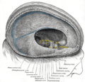 Dura mater and its processes exposed by removing part of the right half of the skull, and the brain.
 -
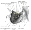
 Superficial dissection of brain-stem. Ventral view.
 -
 Hind- and mid-brains; postero-lateral view.
 -
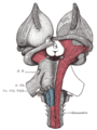
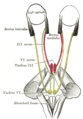 Figure showing the mode of innervation of the Recti medialis and lateralis of the eye.
 -
 Dissection showing origins of right ocular muscles, and nerves entering by the superior orbital fissure.
 -
 Cerebrum.Inferior view.Deep dissection


External links
- hier-541 at NeuroNames
- MedEd at Loyola GrossAnatomy/h_n/cn/cn1/cn6.htm
- Animations of extraocular cranial nerve and muscle function and damage (University of Liverpool)
- cranialnerves at The Anatomy Lesson by Wesley Norman (Georgetown University) (VI)