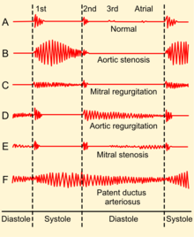
Phonocardiogram
| Phonocardiogram | |
|---|---|
| Synonyms | PCG |
| ICD-9-CM | 89.55 |

A phonocardiogram (or PCG) is a plot of high-fidelity recording of the sounds and murmurs made by the heart with the help of the machine called the phonocardiograph; thus, phonocardiography is the recording of all the sounds made by the heart during a cardiac cycle.[2][3]
Medical use

Heart sounds result from vibrations created by the closure of the
Discrete and the packet wavelet transform
According to a review by Cherif et al.,
History

Awareness of the sounds made by the heart dates to ancient times. The idea of developing an instrument to record it may date back to Robert Hooke (1635–1703), who wrote: "There may also be a possibility of discovering the internal motions and actions of bodies - whether animal, vegetable, or mineral, by the sound they make". The earliest known examples of phonocardiography date to the 1800s.[7]
Monitoring and recording equipment for phonocardiography was developed through the 1930s and 1940s. Standardization began by 1950, when the first international conference was held in Paris.[7]
A phonocardiogram system manufactured by Beckman Instruments was used on at least one of the Project Gemini manned spaceflights (1965-1966) to monitor the heartbeat of astronauts on the flight. It was one of many Beckman Instruments specialized for and used by NASA.[8]
John Keefer filed a patent for a phonocardiogram simulator in 1970 while he was an employee of the U.S. government. The original patent description indicates that it is a device which via electrical voltage mimics the human heart's sounds.[9]
Fetal Phonocardiogram
A fetal phonocardiogram (or fPCG) is a specialized application of phonocardiography designed to be a non-invasive diagnostic technique to capture the sounds of the fetal heart in utero. These fetal phonocardiograms can be analyzed to detect any abnormalities in the fetal heart. Fetal phonocardiography has become an important tool in prenatal care, as it allows clinicians to detect and monitor potential heart problems in the fetus before birth.[10]
The use of phonocardiography to study the fetal heart dates back to the 1960s, when researchers first began to explore the feasibility of detecting fetal heart sounds using external microphones.[10] Early studies focused on using phonocardiography to measure fetal heart rate and rhythm. Over time, advances in technology and techniques have enabled researchers to use fetal phonocardiography to detect a wider range of fetal heart abnormalities.[11][12] Fetal phonocardiography is typically performed during routine prenatal visits, starting around 18–20 weeks of gestation. The procedure involves placing a small microphone on the mother's abdomen over the fetal heart. The microphone captures the sounds of the fetal heart, which are then amplified and recorded for analysis. Khandoker et al. developed a multi-channel fetal phonocardiogram (fPCG) with four sound transducers applied in a simple and consistent pattern across the maternal abdomen.[13][14] The intellectual property (IP) technology license was given to the home-based monitoring device, the Emirati startup, that helps pregnant mothers monitor fetal heartbeat and the baby’s cardiac activity.[15]
See also
- EKG
- Echocardiogram
References
- ^ https://www.ncbi.nlm.nih.gov/bookshelf/br.fcgi?book=cm&part=A622&rendertype=figure&id=A633 Chapter 8/no page given/Google
- PMID 26871603.
- ISBN 9781447107736.Chapter 8/no Google page given
- ISBN 9780323375238.
- ISBN 978-1418020668. Retrieved 27 November 2016.
phonocardiogram purpose.
- .
- ^ ISSN 0097-1049.
- ^ "Beckman Instruments Supplying Medical Flight Monitoring Equipment" (PDF). Space News Roundup. March 3, 1965. pp. 4–5. Retrieved 7 August 2019.
- ^ M, Keefer John (Apr 28, 1970), Phonocardiogram simulator, retrieved 2016-06-02
- ^ ISBN 978-0-407-00450-4, retrieved 2023-08-30
- PMID 28944222.
- S2CID 32510704.
- PMID 30206289.
- ^ "Novel digital device to detect heart murmurs". gulfnews.com. 2015-03-28. Retrieved 2023-08-30.
- ^ National, The (2020-03-18). "Khalifa University grants IP licence to Emirati startup". The National. Retrieved 2023-08-30.
Further reading
- Almasi, Ali; Shamsollahi, Mohammad-Bagher; Senhadji, Lotfi (2011-08-01). "A dynamical model for generating synthetic Phonocardiogram signals". 2011 Annual International Conference of the IEEE Engineering in Medicine and Biology Society. Vol. 2011. pp. 5686–5689. PMID 22255630.
- Mizuno, Atsushi; Niwa, Koichiro; Shirai, Takeaki; Shiina, Yumi (1 January 2015). "Phonocardiogram in adult patients with tetralogy of Fallot". Journal of Cardiology. 65 (1): 82–86. PMID 24842232. Retrieved 2 June 2016.
- Chernecky, Cynthia C.; Berger, Barbara J. (2007). Laboratory Tests and Diagnostic Procedures. Elsevier Health Sciences. ISBN 978-1416066835. Retrieved 27 November 2016.
