
Submasseteric space
| Submasseteric space | |
|---|---|
 Location of the masseter muscles. The submasseteric space is between the masseter and the mandible. | |
 The four compartents of the right masticator space. A Temporalis muscle, B Masseter muscle, C Lateral pterygoid muscle, D Medial ptaerygoid muscle, E Superficial temporal space, F Deep temporal space, G Submasseteric space, H Pterygomandibular space, I Approximate location of infratemporal space | |
| Anatomical terminology |
The submasseterric space (also termed the masseteric space) is a
Structure
Anatomic boundaries
The boundaries of each submasseteric space are:
- the anterior margin of the masseter muscle anteriorly,
- the parotid gland posteriorly,[1]
- the zygomatic arch superiorly,[1]
- the inferior border of the mandible inferiorly,[1]
- the lateral surface of the mandibular ramus medially (the submasseteric space is superficial to the mandible),[1]
- the masseter muscle laterally (the submasseteric space is deep to masseter).[1]
Communications
The communications of each submasseteric space are:
- to the buccal space anteriorly,
- to the pterygomandibular space (around the posterior margin of the mandibular ramus to its medial surface) and the parotid space posteriorly,
- to the superficial temporal space superiorly.
Contents
In health, the space contains:
- the masseteric artery and vein.[1]
Clinical significance
Submasseteric abscesses are relatively rare, and may be confused with a parotid abscess or parotitis.[2] They tend to be chronic.[3] The submasseteric space may be involved by infections that spread from the buccal space.[1] Sometimes mandibular fractures in the region of the angle of the mandible may cause an infection of the submasseteric space.[1] The signs and symptoms of a submasseteric abscess may include marked trismus (i.e. difficulty opening the mouth, since the masseter elevates the mandible and it becomes restricted) and swelling in the region of the masseter muscle.[1] The treatment of a submasseteric space infection is usually by surgical incision and drainage, and the incision is placed intra-orally (inside the mouth) or both intra and extra-orally if other parts of the masticator space are involved.
Odontogenic infections
The submasseteric space is sometimes involved by the spread of
Additional images
-
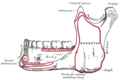 Diagram showing lateral surface of the mandible and the area of insertion of the masseter muscle on the mandibular ramus.
 -
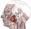 The left masseter muscle (red highlight), shown partially covered by superficial muscles such as the platysma muscle, the zygomaticus major muscle and the zygomaticus minor muscle

.png)
References
- ^ ISBN 9780323049030.
- PMID 21525699.
- PMID 12812946.
- ISBN 978-0-323-06489-7.)
{{cite book}}:|last=has generic name (help)CS1 maint: multiple names: authors list (link