Tetrachromacy
Tetrachromacy (from Ancient Greek tetra, meaning "four" and chroma, meaning "color") is the condition of possessing four independent channels for conveying color information, or possessing four types of cone cell in the eye. Organisms with tetrachromacy are called tetrachromats.
In tetrachromatic organisms, the sensory color space is four-dimensional, meaning that matching the sensory effect of arbitrarily chosen spectra of light within their visible spectrum requires mixtures of at least four primary colors.
Tetrachromacy is demonstrated among several species of birds,[2] fishes,[3] and reptiles.[3] The common ancestor of all vertebrates was a tetrachromat, but a common ancestor of mammals lost two of its four kinds of cone cell, evolving dichromacy, a loss ascribed to the conjectured nocturnal bottleneck. Some primates then later evolved a third cone.[4]
Physiology
The normal explanation of tetrachromacy is that the organism's
Humans

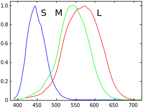
Tetrachromacy requires that there be four independent photoreceptor cell classes with different spectral sensitivity. However, there must also be the appropriate post-receptoral mechanism to compare the signals from the four classes of receptors. According to the opponent process theory, humans have three opponent channels, which give trichromacy. It is unclear whether having available a fourth opponent channel is sufficient for tetrachromacy.[citation needed]
Mice, which normally have only two cone pigments (and therefore two opponent channels), have been engineered to express a third cone pigment, and appear to demonstrate increased chromatic discrimination,[7] possibly indicating trichromacy, and suggesting they were able to create or re-enable a third opponent channel. This would support the theory that humans should be able to utilize a fourth opponent channel for tetrachromatic vision. However, the original publication's claims about plasticity in the optic nerve have also been disputed.[8]
Tetrachromacy in carriers of CVD
It has been theorized that females who carry
In humans, two
Variation in cone pigment genes is widespread in most human populations, but the most prevalent and pronounced tetrachromacy would derive from female carriers of major red / green pigment anomalies, usually classed as forms of "
In humans, preliminary visual processing occurs in the neurons of the retina. It is not known how these nerves would respond to a new color channel: Whether they would handle it separately, or just combine it with one of the existing channels. Similarly, visual information leaves the eye by way of the optic nerve, and a variety of final image processing takes place in the brain; it is not known whether the optic nerve or the areas of the brain have any capacity to effectively respond if presented with a stimulus from a new color signal.
Tetrachromacy may also enhance vision in dim lighting, or in looking at a screen.[14][failed verification]
Conditional tetrachromacy
Despite being trichromats, humans can experience slight tetrachromacy at low light intensities, using their mesopic vision. In mesopic vision, both cone cells and rod cells are active. While rods typically do not contribute to color vision, in these specific light conditions, they may give a small region of tetrachromacy in the color space.[15] Human rod cell sensitivity is greatest at 500 nm (bluish-green) wavelength, which is significantly different from the peak spectral sensitivity of the cones (typically 420, 530, and 560 nm).
Blocked tetrachromacy
Although many birds are tetrachromats with a fourth color in the ultraviolet, humans cannot see ultraviolet light directly because the
While an extended visible range does not denote tetrachromacy, some believe that visual pigments are available with sensitivity in
However, there is no peer-reviewed evidence supporting this claim.Other animals

Fish
Fish, specifically teleosts, are typically tetrachromats.[3] Exceptions include:
- Sharks and rays – range from monochromacy to trichromacy[3]
- Deep-sea fish – often rod monochromats
- pentachromacy or higher[3]
Birds
Some species of birds, such as the
The four cone types, and the specialization of pigmented oil droplets, give birds better color vision than that of humans.
Some birds such as
Pentachromacy and greater
The dimensionality of color vision has no upper bound, but vertebrates with color vision greater than tetrachromacy are rare. The next level is pentachromacy, which is five-dimensional color vision requiring at least 5 different classes of photoreceptor as well as 5 independent channels of color information through the primary visual system.
A female that is heterozygous for both the
Some
Invertebrates can have large numbers of different opsin classes, including 15 opsins in bluebottle butterflies[29] or 33 in mantis shrimp.[30] However, it has not been shown that color vision in these invertebrates is of a dimension commensurate with the number of opsins.
See also
References
- S2CID 19458550.
- ^ Goldsmith, Timothy H. (2006). "What Birds See". Scientific American (July 2006): 69–75.
- ^ S2CID 52808112.
- PMID 19720656.
- ISBN 9783110806984.)
{{cite book}}: CS1 maint: multiple names: authors list (link - ^ a b
Jordan, Gabriele; Deeb, Samir S.; Bosten, Jenny M.; Mollon, J.D. (July 2010). "The dimensionality of color vision in carriers of anomalous trichromacy". Journal of Vision. 10 (12): 12. PMID 20884587.
- ^
Jacobs, Gerald H.; Williams, Gary A.; Cahill, Hugh; Nathans, Jeremy (23 March 2007). "Emergence of novel color vision in mice engineered to express a human cone photopigment". S2CID 85273369.
- ^
Makous, W. (12 October 2007). "Comment on 'Emergence of novel color vision in mice engineered to express a human cone photopigment' ". Science. 318 (5848): 196. PMID 17932271.
- ^ Jameson, K.A.; Highnote, S.M.; Wasserman, L.M. (2001). "Richer color experience in observers with multiple photopigment opsin genes" (PDF). .
- ^
Jordan, G. (July 1993). "A study of women heterozygous for colour deficiencies". Vision Research. 33 (11): 1495–1508. S2CID 17648762.
- ^
Backhaus, Werner G.K.; Backhaus, Werner; Kliegl, Reinhold; Werner, John Simon (1998). Color Vision: Perspectives from different disciplines. Walter de Gruyter. ISBN 9783110161007– via Google books.
- ^ San Diego woman, Concetta Antico, diagnosed with 'super vision' (video). 22 Nov 2013 – via YouTube.
- ^
Francis, Richard C. (2011). "Chapter 8. X-Women". Epigenetics: The ultimate mystery of inheritance. New York, NY & London, UK: W. W. Norton. pp. 93–104. ISBN 978-0-393-07005-7.
- ^ Robson, David (September 5, 2014). "The Women with Superhuman Vision". BBC News. Archived from the original on September 13, 2014. Retrieved December 30, 2017.
- ^
Autrum, Hansjochem & Jung, Richard (1973). Integrative Functions and Comparative Data. Vol. 7. Springer-Verlag. p. 226. ISBN 978-0-387-05769-9– via Google books.
- ^
Mainster, M.A. (2006). "Violet and blue light blocking intraocular lenses: Photoprotection versus photoreception". PMID 16714268.
- ^ Hambling, David (29 May 2002). "Let the light shine in". The Guardian.
- ^ Fulton, James T. (31 July 2009). "The human is a blocked tetrachromat". neuronresearch.net. Retrieved 1 June 2022.
- S2CID 4347875.
- S2CID 2484928.
- ISBN 978-0-12-004529-7.
- S2CID 372159.
- PMID 25609782.
- ^ "Crow curiosities: can crows see UV?". Corvid Research. 2 December 2020. Retrieved 2 November 2024.
- ^ S2CID 3138290. Retrieved 13 June 2023.
- ISSN 1084-9521.
- S2CID 12462107.
- S2CID 5932623.
- .
- ^ Hansen, Sarah (17 July 2020). "Mantis Shrimp Eyes Get Even Wilder: UMBC Team Finds Twice The Expected Number Of Light-detecting Proteins - UMBC: University Of Maryland, Baltimore County". UMBC. Retrieved 7 October 2022.
External links
- Goldsmith, Timothy H. "What Birds See" Scientific American July 2006. An article about the tetrachromatic vision of birds
- Thompson, Evan (2000). "Comparative color vision: Quality space and visual ecology." In Steven Davis (Ed.), Color Perception: Philosophical, Psychological, Artistic and Computational Perspectives, pp. 163–186. Oxford: Oxford University Press.
- Looking for Madam Tetrachromat By Glenn Zorpette. Red Herring magazine, 1 November 2000
- "Exploring the fourth dimension" Archived 2016-04-15 at the Wayback Machine. University of Bristol School of Biological Sciences. March 20, 2009.
- Colors - The Perfect Yellow By Radiolab, 21 May 2012 (Explores tetrachromacy in humans)
- The dimensionality of color vision in carriers of anomalous trichromacy--Gabriele Jordan et al--Journal of Vision August 12, 2010:
- On Tetrachromacy Ágnes Holba & B. Lukács

