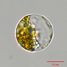
Thalassiosira pseudonana
| Thalassiosira pseudonana | |
|---|---|

| |
| A Thalassiosira pseudonana oogonium (egg cell) beginning to expand through the cell wall. Artificial coloring denotes chlorophyll (blue) and DNA (red). | |
| Scientific classification | |
| Domain: | Eukaryota |
| Clade: | Diaphoretickes |
| Clade: | SAR |
| Clade: | Stramenopiles |
| Phylum: | Gyrista |
| Subphylum: | Ochrophytina |
| Class: | Bacillariophyceae |
| Order: | Thalassiosirales |
| Family: | Thalassiosiraceae |
| Genus: | Thalassiosira |
| Species: | T. pseudonana
|
| Binomial name | |
| Thalassiosira pseudonana Hasle & Heimdal, 1970
| |
Thalassiosira pseudonana is a species of marine centric
The
Morphology

Thalassiosira pseudonana has a
Biomineralization
The distinct nano- to micro-scale structure of T. pseudonana follows a specific mechanism of formation. It begins with the formation of a thin base layer that outlines the valve.[7] Next, there is radiation of the silica ribs and the buildup of the rims, while portulae develop. The formation of the radial structure starts from a central location and spreads out in x and y axes planes.[7] During maturation, the valve surface becomes increasingly silicified, and the rim continues to develop, while the central portion of the valve becomes more rigid. Initially, the valve nanopores have irregular shapes, but they become circular during maturation.[7] The nanoscale structures of T. pseudonana are genetically mediated. The silicification process involves three categories of molecules: silaffins, which are highly post-translationally modified phosphoproteins; long chain polyamines (LCPAs); and silacidins, which are acidic proteins.[4] During valve synthesis, mRNA levels for silaffin 3 increase and lead to the formation of the base layer.[7] The presence of lower concentrations of silaffin 3 or the light form of silaffin 1 and 2 leads to the generation of spherical silica structures, indicating possible mechanisms involved in the formation of spherical silica in the ridges.[7] Over 150 genes have been identified as playing a role in the silicon biomineralization of T. pseudonana. A set of 75 genes were upregulated only during silicon limitation, while 84 genes were upregulated by both silicon and iron limitations, indicating a linkage between their iron and silicon pathways.[4][10] T. pseudonana also possesses chitin-based scaffolds that are important in the formation of their biosilica structure.[11]
Symbiosis
Thalassiosira pseudonana and the heterotrophic alphaproteobacterium Ruegeria pomeroyi form a chemical symbiosis in coculture. The bacteria provide vitamin B12 to the diatoms, which in exchange provide organic nutrients to the bacteria. In the presence of the diatom, the bacteria start producing a transporter for dihydroxypropanesulfonate (DHPS), a nutrient produced by the diatom for the bacteria.[12] A metabolic survey of the association between the bacterium Dinoroseobacter shibae and T. pseudonana showed that the bacterium has minimal impact on the growth of T. pseudonana, but it causes metabolic changes by upregulating the intracellular amino acids and amino acid derivatives of the diatom.[13] It has been demonstrated that under conditions of environmental instability and extreme warming, biofilm formation can accelerate the evolutionary responses of T. pseudonana.[14]
References
- S2CID 8593895.
- ^ Clement, R., Dimnet, L., Maberly, S. C., Gontero, B. 2015. The nature of the CO2‐concentrating mechanisms in a marine diatom, Thalassiosira pseudonana. New Phytologist Trust 209: 1417–1427.
- PMID 27827427.
- ^ S2CID 3738371.
- ^ S2CID 85688848.
- ^ a b Rad Menendez, Cecilia (2011). Phenotypic and genotypic characterization of Thalassiosira pseudonana (Bacillariophyceae) strains (Ph.D. thesis). University of Aberdeen.
- ^ S2CID 138652614.
- ^ ISSN 0967-0262.
- S2CID 4336989.
- PMID 18212125.)
{{cite journal}}: CS1 maint: multiple names: authors list (link - S2CID 23179914.
- .
- S2CID 255127394.
- S2CID 92224508.
Further reading
Meksiarun, Phiranuphon; Spegazzini, Nicolas; Matsui, Hiroaki; Nakajima, Kensuke; Matsuda, Yusuke; Sato, Hidetoshi (January 2015). "In Vivo Study of Lipid Accumulation in the Microalgae Marine Diatom Thalassiosira pseudonana Using Raman Spectroscopy". Applied Spectroscopy. 69 (1): 45–51.
Yi, Andy Xianliang; Leung, Priscilla T. Y.; Leung, Kenneth M. Y. (September 2014). "Photosynthetic and molecular responses of the marine diatom Thalassiosira pseudonana to triphenyltin exposure". Aquatic Toxicology. 154: 48–57.
Luo, Chun-Shan; Liang, Jun-Rong; Lin, Qun; Li, Caixia; Bowler, Chris; Anderson, Donald; Wang, Peng; Wang, Xin-Wei; Gao, Ya-Hui (December 2014). "Cellular Responses Associated with ROS Production and Cell Fate Decision in Early Stress Response to Iron Limitation in the Diatom Thalassiosira pseudonana". Journal of Proteome Research. 13 (12): 5510–5523.
Sunda, William G.; Shertzer, Kyle W.; Coggins, Lew (October 2014). "Positive feedbacks between bottom-up and top-down controls promote the formation and toxicity of ecosystem disruptive algal blooms: A modeling study". Harmful Algae. 39: 342–356. .
Samukawa, Mio; Shen, Chen; Hopkinson, Brian; Matsuda, Yusuke (2014). "Localization of putative carbonic anhydrases in the marine diatom, Thalassiosira pseudonana". Photosynthesis Research. 121 (2–3): 235–249.
Delalat, Bahman; Sheppard, Vonda C.; Ghaemi, Soraya Rasi; Rao, Shasha; Prestidge, Clive A.; McPhee, Gordon; Rogers, Mary-Louise; Donoghue, Jaqueline F.; Pillay, Vinochani; Johns, Terrance G.; Kroeger, Nils; Voelcker, Nicolas H. (2015). "Targeted drug delivery using genetically engineered diatom biosilica". Nature Communications. 6: 8791.

