
Sarcoidosis
| Sarcoidosis | |
|---|---|
| Other names | Sarcoïdosis, sarcoid, Besnier–Boeck–Schaumann disease pulmonary eosinophilia[7] |
| Treatment | Ibuprofen, prednisone, methotrexate[8][9] |
| Prognosis | Mortality 1–7%[5] |
| Frequency | 1.9 million with interstitial lung disease (2015)[10] |
| Deaths | 122,000 with interstitial lung disease (2015)[11] |
Sarcoidosis (also known as Besnier–Boeck–Schaumann disease) is a disease involving abnormal collections of inflammatory cells that form lumps known as
The cause of sarcoidosis is unknown.
Sarcoidosis may resolve without any treatment within a few years.[2][5] However, some people may have long-term or severe disease.[5] Some symptoms may be improved with the use of anti-inflammatory drugs such as ibuprofen.[8] In cases where the condition causes significant health problems, steroids such as prednisone are indicated.[9] Medications such as methotrexate, chloroquine, or azathioprine may occasionally be used in an effort to decrease the side effects of steroids.[9] The risk of death is 1–7%.[5] The chance of the disease returning in someone who has had it previously is less than 5%.[2]
In 2015, pulmonary sarcoidosis and interstitial lung disease affected 1.9 million people globally and they resulted in 122,000 deaths.[10][11] It is most common in Scandinavians, but occurs in all parts of the world.[14] In the United States, risk is greater among black people as opposed to white people.[14] It usually begins between the ages of 20 and 50.[4] It occurs more often in women than men.[4] Sarcoidosis was first described in 1877 by the English doctor Jonathan Hutchinson as a non-painful skin disease.[15]
Signs and symptoms


Sarcoidosis is a systemic inflammatory disease that can affect any organ, although it can be
The combination of erythema nodosum, bilateral hilar lymphadenopathy, and joint pain is called Löfgren syndrome, which has a relatively good prognosis.[20] This form of the disease occurs significantly more often in Scandinavian patients than in those of non-Scandinavian origin.[26]
Respiratory tract
Localization to the lungs is by far the most common manifestation of sarcoidosis.
The four stages of pulmonary involvement are based on radiological stage of the disease, which is helpful in prognosis:[32]
- Stage I: bilateral hilar lymphadenopathy (BHL) alone
- Stage II: BHL with pulmonary infiltrates
- Stage III: pulmonary infiltrates without BHL
- Stage IV: fibrosis
Use of the Scadding scale only provides general information regarding the prognosis of the pulmonary disease over time. Caution is recommended, as it only shows a general relation with physiological markers of the disease and the variation is such that it has limited applicability in individual assessments, including treatment decisions.[12]
Skin
Sarcoidosis involves the skin in between 9 and 37% of cases and is more common in
Heart
Histologically, sarcoidosis of the heart is an active granulomatous inflammation surrounded by reactive oedema. The distribution of affected areas is patchy with localised enlargement of heart muscles. This causes scarring and remodelling of the heart, which leads to dilatation of heart cavities and thinning of heart muscles. As the situation progresses, it leads to
The frequency of cardiac involvement varies and is significantly influenced by race; in Japan, more than 25% of those with sarcoidosis have symptomatic cardiac involvement, whereas in the US and Europe, only about 5% of cases present with cardiac involvement.[28] Autopsy studies in the US have revealed a frequency of cardiac involvement of about 20–30%, whereas autopsy studies in Japan have shown a frequency of 60%.[22] The presentation of cardiac sarcoidosis can range from asymptomatic conduction abnormalities to fatal ventricular arrhythmia.[39][40]
Conduction abnormalities are the most common cardiac manifestations of sarcoidosis in humans and can include complete
Eye
Eye involvement occurs in about 10–90% of cases.[22] Manifestations in the eye include uveitis, uveoparotitis, and retinal inflammation, which may result in loss of visual acuity or blindness.[50] The most common ophthalmologic manifestation of sarcoidosis is uveitis.[22][51][52] The combination of anterior uveitis, parotitis, VII cranial nerve paralysis and fever is called uveoparotid fever or Heerfordt syndrome (D86.8). Development of scleral nodule associated with sarcoidosis has been observed.[53]
Nervous system
Any of the components of the nervous system can be involved.[54] Sarcoidosis affecting the nervous system is known as neurosarcoidosis.[54] Cranial nerves are most commonly affected, accounting for about 5–30% of neurosarcoidosis cases, and peripheral facial nerve palsy, often bilateral, is the most common neurological manifestation of sarcoidosis.[54][55][56] It occurs suddenly and is usually transient. The central nervous system involvement is present in 10–25% of sarcoidosis cases.[31] Other common manifestations of neurosarcoidosis include optic nerve dysfunction, papilledema, palate dysfunction, neuroendocrine changes, hearing abnormalities, hypothalamic and pituitary abnormalities, chronic meningitis, and peripheral neuropathy.[28] Myelopathy, that is spinal cord involvement, occurs in about 16–43% of neurosarcoidosis cases and is often associated with the poorest prognosis of the neurosarcoidosis subtypes.[54] Whereas facial nerve palsies and acute meningitis due to sarcoidosis tend to have the most favourable prognosis,[54] another common finding in sarcoidosis with neurological involvement is autonomic or sensory small-fiber neuropathy.[57][58] Neuroendocrine sarcoidosis accounts for about 5–10% of neurosarcoidosis cases and can lead to diabetes insipidus, changes in menstrual cycle and hypothalamic dysfunction.[54][56] The latter can lead to changes in body temperature, mood, and prolactin (see the endocrine and exocrine section for details).[54]
Endocrine and exocrine
Thyroid dysfunction is seen in 4.2–4.6% of cases.[62][63]
Gastrointestinal and genitourinary
Symptomatic gastrointestinal (GI) involvement occurs in less than 1% of cases (if one excludes the liver), and most commonly the stomach is affected, although the small or large intestine may also be affected in a small portion of cases.
Around 70% of people have granulomas in their livers, although only in about 20–30% of cases, liver function test anomalies reflecting this fact are seen.
Blood
Abnormal blood tests are frequent, accounting for over 50% of cases, but are not diagnostic.
Lymphadenopathy (swollen glands) is common in sarcoidosis and occurs in 15% of cases.[23] Intrathoracic nodes are enlarged in 75 to 90% of all people; usually this involves the hilar nodes, but the paratracheal nodes are commonly involved. Peripheral lymphadenopathy is very common, particularly involving the cervical (the most common head and neck manifestation of the disease), axillary, epitrochlear, and inguinal nodes.[71] Approximately 75% of cases show microscopic involvement of the spleen, although only in about 5–10% of cases does splenomegaly appear.[19][64]
Bone, joints, and muscles
Sarcoidosis can be involved with the joints, bones, and muscles. This causes a wide variety of musculoskeletal complaints that act through different mechanisms.[72] About 5–15% of cases affect the bones, joints, or muscles.[31]
Arthritic syndromes can be categorized as acute or chronic.[72] Sarcoidosis patients with acute arthritis often also have bilateral
Chronic sarcoid arthritis usually occurs in the setting of more diffuse organ involvement.[72] The ankles, knees, wrists, elbows, and hands may all be affected in the chronic form and often this presents itself in a polyarticular pattern.[72] Dactylitis similar to that seen in psoriatic arthritis, that is associated with pain, swelling, overlying skin erythema, and underlying bony changes may also occur.[72] Development of Jaccoud arthropathy (a nonerosive deformity) is very rarely seen.[72]
Bone involvement in sarcoidosis has been reported in 1–13% of cases.[56] The most frequent sites of involvement are the hands and feet, whereas the spine is less commonly affected.[72] Half of the patients with bony lesions experience pain and stiffness, whereas the other half remain asymptomatic.[72] Periostitis is rarely seen in sarcoidosis and has been found to present itself at the femoral bone.[73][74]
Cause
The exact cause of sarcoidosis is not known.[2] The current working hypothesis is, in genetically susceptible individuals, sarcoidosis is caused through alteration to the immune response after exposure to an environmental, occupational, or infectious agent.[75] Some cases may be caused by treatment with tumor necrosis factor (TNF) inhibitors like etanercept.[76]
Genetics
The heritability of sarcoidosis varies according to
Investigations of genetic susceptibility yielded many candidate genes, but only few were confirmed by further investigations and no reliable genetic markers are known. Currently, the most interesting candidate gene is
Infectious agents
Several infectious agents appear to be significantly associated with sarcoidosis, but none of the known associations is specific enough to suggest a direct causative role.
Autoimmune
Association of
Pathophysiology
While TNF is widely believed to play an important role in the formation of granulomas (this is further supported by the finding that in animal models of mycobacterial granuloma formation inhibition of either TNF or IFN-γ production inhibits granuloma formation), sarcoidosis can and does still develop in those being treated with TNF antagonists like etanercept.[94]
B cells also likely play a role in the pathophysiology of sarcoidosis.
Cases of sarcoidosis have also been reported as part of the
Histopathology
Sarcoidosis is characterized by the formation of non-necrotizing ("non-caseating")
-
 Sarcoidosis in a lymph node
 -
 asteroid bodies
 -
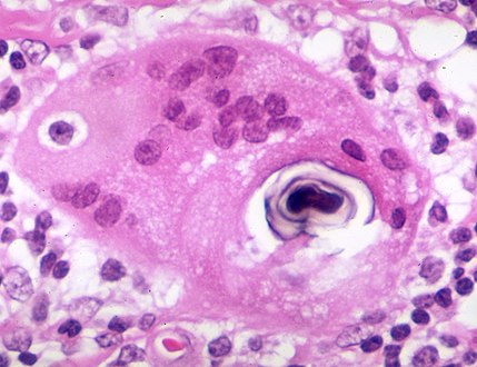 Schaumann body in sarcoidosis
 -
 Asteroid body in sarcoidosis
 -
 Hamazaki–Wesenberg bodies in sarcoidosis in lymph node
_lymph_node_biopsy.jpg)


.jpg)
_bodies-_Lymph_node_(6134890353).jpg)
Diagnosis
Diagnosis of sarcoidosis is a
Serum markers of sarcoidosis, include:
Differential diagnosis includes metastatic disease, lymphoma, septic emboli,
Chest radiograph changes are divided into four stages:[104]
- bihilar lymphadenopathy
- bihilar lymphadenopathy and reticulonodular infiltrates
- bilateral pulmonary infiltrates
- fibrocystic sarcoidosis typically with upward hilar retraction, cystic and bullous changes
Although people with stage 1 radiographs tend to have the acute or subacute, reversible form of the disease, those with stages 2 and 3 often have the chronic, progressive disease; these patterns do not represent consecutive "stages" of sarcoidosis. Thus, except for epidemiologic purposes, this categorization is mostly of historic interest.[28]
In sarcoidosis presenting in the Caucasian population, hilar adenopathy and erythema nodosum are the most common initial symptoms. In this population, a biopsy of the gastrocnemius muscle is a useful tool in correctly diagnosing the person. The presence of a noncaseating epithelioid granuloma in a gastrocnemius specimen is definitive evidence of sarcoidosis, as other tuberculoid and fungal diseases extremely rarely present histologically in this muscle.[105]
PET scan is able to quantify disease activity which cannot be performed by CMR.[107]
-
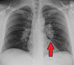 Hilar adenopathy especially on the person's left (AP CXR)
 -
 Hilar adenopathy especially on the person's left (lateral CXR)
 -
 Hilar adenopathy especially on the person's left (coronal CT)
 -
 Hilar adenopathy especially on the person's left (transverse CT)


Classification
Sarcoidosis may be divided into the following types:[36]
- Annular sarcoidosis
- Erythrodermic sarcoidosis
- Ichthyosiform sarcoidosis
- Hypopigmented sarcoidosis
- Löfgren syndrome
- Lupus pernio
- Morpheaform sarcoidosis
- Mucosal sarcoidosis
- Neurosarcoidosis
- Papular sarcoid
- Scar sarcoid
- Subcutaneous sarcoidosis
- Systemic sarcoidosis
- Ulcerative sarcoidosis
Treatment
Treatments for sarcoidosis vary greatly depending on the patient.[108] At least half of patients require no systemic therapy.[109] Most people (>75%) only require symptomatic treatment with nonsteroidal anti-inflammatory drugs (NSAIDs) like ibuprofen or aspirin.[110] For those presenting with lung symptoms, unless the respiratory impairment is devastating, active pulmonary sarcoidosis is observed usually without therapy for two to three months; if the inflammation does not subside spontaneously, therapy is instituted.[28]
Major categories of drug interventions include
Corticosteroids, most commonly prednisone or prednisolone, have been the standard treatment for many years.[19] In some people, this treatment can slow or reverse the course of the disease, but other people do not respond to steroid therapy. The use of corticosteroids in mild disease is controversial because in many cases the disease remits spontaneously.[111][112]
Antimetabolites
Antimetabolites, also categorized as
Immunosuppressants
As the granulomas are caused by collections of immune system cells, particularly
In a clinical trial cyclosporine added to prednisone treatment failed to demonstrate any significant benefit over prednisone alone in people with pulmonary sarcoidosis, although there was evidence of increased toxicity from the addition of cyclosporine to the steroid treatment including infections, malignancies (cancers), hypertension, and kidney dysfunction.[116] Likewise chlorambucil and cyclophosphamide are seldom used in the treatment of sarcoidosis due to their high degree of toxicity, especially their potential for causing malignancies.[127] Infliximab has been used successfully to treat pulmonary sarcoidosis in clinical trials in a number of cases.[116] Etanercept, on the other hand, has failed to demonstrate any significant efficacy in people with uveal sarcoidosis in a couple of clinical trials.[116] Likewise golimumab has failed to show any benefit in those with pulmonary sarcoidosis.[116] One clinical trial of adalimumab found treatment response in about half of subjects, which is similar to that seen with infliximab, but as adalimumab has better tolerability profile it may be preferred over infliximab.[116]
Specific organ treatments
Recently selective
Because of its uncommon nature, the treatment of male reproductive tract sarcoidosis is controversial. Since the differential diagnosis includes testicular cancer, some recommend orchiectomy, even if evidence of sarcoidosis in other organs is present. In the newer approach, testicular, epididymal biopsy and resection of the largest lesion has been proposed.[67]
Symptoms
People with sarcoidosis may have a range of symptoms that do not correspond with objective physical evidence of disease but that still decrease quality of life.[138]
Physical therapy, rehabilitation, and counseling can help avoid deconditioning,[138]: 733 and improve social participation, psychological well-being, and activity levels. Key aspects are avoiding exercise intolerance and muscle weakness.[138]: 734
Low or moderate-intensity physical training has been shown to improve fatigue, psychological health, and physical functioning in people sarcoidosis without adverse effects.[139][140] Inspiratory muscle training has also decreased severe fatigue perception in subjects with early stages of sarcoidosis, as well as improving functional and maximal exercise capacity and respiratory muscle strength.[141] The duration, frequency, and physical intensity of exercise needs to accommodate impairments such as joint pain, muscle pain, and fatigue.[138]: 734 [140][142]
Neurostimulants such as methylphenidate and modafinil have shown some effectiveness as an adjunct for treatment of sarcoidosis fatigue.[138]: 733 [143]
Treatments for symptomatic neuropathic pain in sarcoidosis patients is similar to that for other causes, and include antidepressants, anticonvulsants and prolonged-release opioids, however, only 30 to 60% of patients experience limited pain relief.[138]: 733
Prognosis

The disease can remit spontaneously or become chronic, with exacerbations and remissions. In some cases, it can progress to pulmonary fibrosis and death. In benign cases, remission can occur in 24 to 36 months without treatment but regular follow ups are required. Some cases, however, may persist several decades.[19] Two-thirds of people with the condition achieve a remission within 10 years of the diagnosis.[144] When the heart is involved, the prognosis is generally less favourable, though corticosteroids appear effective in improving AV conduction.[145][146] The prognosis tends to be less favourable in African Americans than in white Americans.[26] In a Swedish population-based analysis, the majority of cases who did not have severe disease at diagnosis had comparable mortality to the general population.[147] The risk for premature death was markedly (2.3-fold) increased compared to the general population for a smaller group of cases with severe disease at diagnosis.[147] Serious infections, sometimes multiple during the course of disease, and heart failure might contribute to the higher risk of early death in some patients with sarcoidosis.[148][149]
Some 1990s studies indicated that people with sarcoidosis appear to be at significantly increased risk for cancer, in particular
Epidemiology
Sarcoidosis most commonly affects young adults of both sexes, although studies have reported more cases in females. Incidence is highest for individuals younger than 40 and peaks in the age-group from 20 to 29 years; a second peak is observed for women over 50.[19][145]
Sarcoidosis occurs throughout the world in all races with an average incidence of 16.5 per 100,000 in men and 19 per 100,000 in women. The disease is most common in Northern European countries and the highest annual incidence of 60 per 100,000 is found in Sweden and Iceland. In the United Kingdom the prevalence is 16 in 100,000.
There also has been a seasonal clustering observed in sarcoidosis-affected individuals.[167] In Greece about 70% of diagnoses occur between March and May every year, in Spain about 50% of diagnoses occur between April and June, and in Japan it is mostly diagnosed during June and July.[167]
The differing incidence across the world may be at least partially attributable to the lack of screening programs in certain regions of the world, and the overshadowing presence of other granulomatous diseases, such as tuberculosis, that may interfere with the diagnosis of sarcoidosis where they are prevalent.[145] There may also be differences in the severity of the disease between people of different ethnicities. Several studies suggest the presentation in people of African origin may be more severe and disseminated than for Caucasians, who are more likely to have asymptomatic disease.[69] Manifestation appears to be slightly different according to race and sex. Erythema nodosum is far more common in men than in women and in Caucasians than in other races. In Japanese people, ophthalmologic and cardiac involvement are more common than in other races.[19]
It is more common in certain occupations, namely
History
It was first described in 1877 by Dr.
Etymology
The word "sarcoidosis" comes from Greek [σάρκο-] sarco- meaning "flesh", the suffix -(e)ido (from the Greek εἶδος -eidos [usually omitting the initial e in English as the diphthong epsilon-iota in Classic Greek stands for a long "i" = English ee]) meaning "type", " resembles" or "like", and -sis, a common suffix in Greek meaning "condition". Thus the whole word means "a condition that resembles crude flesh". The first cases of sarcoïdosis, which were recognised as a new pathological entity, in Scandinavia, at the end of the 19th century exhibited skin nodules resembling cutaneous sarcomas, hence the name initially given.[citation needed]
Society and culture
The World Association of Sarcoidosis and Other Granulomatous Disorders (WASOG) is an organisation of physicians involved in the diagnosis and treatment of sarcoidosis and related conditions.[171] WASOG publishes the journal Sarcoidosis, Vasculitis and Diffuse Lung Diseases.[172] Additionally, the Foundation for Sarcoidosis Research (FSR) is devoted to supporting research into sarcoidosis and its possible treatments.[173]
There have been concerns that World Trade Center rescue workers are at a heightened risk for sarcoidosis.[174][175]
Comedian and actor Bernie Mac had sarcoidosis. In 2005, he mentioned that the disease was in remission.[176] His death on August 9, 2008, was caused by complications from pneumonia, though Mac's agent states the sarcoidosis was not related to his fatal pneumonia.[177]
Karen "Duff" Duffy, MTV personality and actress, was diagnosed with neurosarcoidosis in 1995.[178]
American football player Reggie White died in 2004, with pulmonary and cardiac sarcoidosis being contributing factors to his fatal heart arrhythmia.[179]
Singer Sean Levert died in 2008 of sarcoidosis complications.[180]
Manning Marable, a professor of public policy at Columbia University, died of pneumonia in 2011, less than a year after undergoing a double lung transplant following a diagnosis of sarcoidosis. In 2012, he was posthumously awarded a Pulitzer Prize in history for his biography "Malcolm X: A Life of Reinvention."
Joseph Rago, Pulitzer Prize-winning writer known for his work at The Wall Street Journal, died of sarcoidosis complications in 2017.[181]
Several historical figures are suspected of having sarcoidosis. In a 2014 letter to the British medical journal The Lancet, it was suggested that the French Revolution leader Maximilien Robespierre may have had sarcoidosis, causing him impairment during his time as head of the Reign of Terror.[182] The symptoms associated with Ludwig van Beethoven's 1827 death have been described as possibly consistent with sarcoidosis.[183] Author Robert Louis Stevenson (1850–1894) had a history of chronic coughs and chest complaints, and sarcoidosis has been suggested as a diagnosis.[184]
Pregnancy
Sarcoidosis generally does not prevent successful pregnancy and delivery; the increase in estrogen levels during pregnancy may even have a slightly beneficial immunomodulatory effect. In most cases, the course of the disease is unaffected by pregnancy, with improvement in a few cases and worsening of symptoms in very few cases, although it is worth noting that a number of the immunosuppressants (such as
References
- ISBN 978-0-08-046012-3.
- ^ a b c d e f g h i j k l m "What Is Sarcoidosis?". NHLBI. June 14, 2013. Archived from the original on 6 April 2016. Retrieved 28 March 2016.
- ^ a b c d "What Are the Signs and Symptoms of Sarcoidosis?". NHLBI. June 14, 2013. Archived from the original on 7 April 2016. Retrieved 29 March 2016.
- ^ a b c d e "Who Is at Risk for Sarcoidosis?". NHLBI. June 14, 2013. Archived from the original on 7 April 2016. Retrieved 28 March 2016.
- ^ PMID 26593147.
- ^ PMID 26593135.
- ISBN 978-0-323-07699-9.
- ^ S2CID 24353355.
- ^ PMID 26611555.
- ^ PMID 27733282.)
{{cite journal}}:|author1=has generic name (help)CS1 maint: numeric names: authors list (link - ^ PMID 27733281.)
{{cite journal}}:|author1=has generic name (help)CS1 maint: numeric names: authors list (link - ^ PMID 21037016.
- ^ "What Causes Sarcoidosis?". NHLBI. June 14, 2013. Archived from the original on 6 April 2016. Retrieved 28 March 2016.
- ^ PMID 26425148.
- ^ (PDF) from the original on 2016-03-04.
- ^ "Lung Diseases: Sarcoidosis: Signs & Symptoms". National Heart, Lung, and Blood Institute. Archived from the original on May 7, 2009. Retrieved May 9, 2009.
- ^ a b Kamangar N, Rohani P, Shorr AF (6 February 2014). Peters SP, Talavera F, Rice TD, Mosenifar Z (eds.). "Sarcoidosis Clinical Presentation". Medscape Reference. WebMD. Archived from the original on 25 February 2014. Retrieved 19 February 2014.
- S2CID 205784334.
- ^ PMID 18021432.
- ^ a b c d King, TE Jr. (March 2008). "Sarcoidosis: Interstitial Lung Diseases: Merck Manual Home Edition". The Merck Manual Home Edition. Merck Sharp & Dohme Corp. Archived from the original on 20 February 2014. Retrieved 19 February 2014.
- PMID 20665396.
- ^ PMID 19857642.
- ^ S2CID 220114548.
- PMID 32476897.
- PMID 18021484.
- ^ a b c d e f g Kamangar N, Rohani P, Shorr AF (6 February 2014). Peters SP, Talavera F, Rice TD, Mosenifar Z (eds.). "Sarcoidosis". Medscape Reference. WebMD. Archived from the original on 10 February 2014. Retrieved 19 February 2014.
- PMID 22579234.
- ^ ISBN 978-0-07174889-6.
- PMID 19857641.
- PMID 22608948.
- ^ S2CID 21345455.
- ^ Kumar and Clark, Clinical Medicine, 8th edition, p. 846.
- ^ PMID 22579238.
- PMID 22000704.
- PMID 19666758.
- ^ ISBN 978-0808923510.
- from the original on 2 March 2014.
- ^ S2CID 205477032.
- PMID 24819193.
- PMID 16415205.
- ^ S2CID 44374668.
- PMID 9363623.
- PMID 5796402.
- S2CID 12132106.
- PMID 26885492.
- ^ "About Sarcoidosis". Stanford University Sarcoidosis Program. Retrieved 2019-08-09.
- S2CID 235076619.
- S2CID 238217835.
- PMID 22291785.
- PMID 22595776.
- PMID 24704868.
- S2CID 10055356.
- PMID 14516826.
- ^ PMID 22595777.
- PMID 11844727.
- ^ PMID 22094184.
- S2CID 6735421.
- PMID 23304492.
- PMID 12897340.
- PMID 6894783.
- ^ Rheumatology Diagnosis & Therapies (2nd ed.). Philadelphia: Lippincott Williams & Wilkins. 2005. p. 342.
- ^ PMID 16899854.
- PMID 23337134.
- ^ ISBN 978-0721601878.
- PMID 24368949.
- S2CID 13481889.
- ^ PMID 21814320.
- S2CID 9363764.
- ^ S2CID 251816742.
- PMID 3033787.
- PMID 16038376.
- ^ PMID 23597964.
- PMID 26320137.
- PMID 10325674.
- PMID 17684288.
- PMID 22527429.
- ^ PMID 29946010.
- PMID 17684289.
- S2CID 23588871.
- PMID 14656748.
- S2CID 260316603.
- PMID 25398810.
- PMID 22461752.
- ^ PMID 22595775.
- PMID 17537780.
- PMID 8711683.
- PMID 21208877. Archived from the original(PDF) on 2014-02-25. Retrieved 2014-02-20.
- PMID 12002380.
- PMID 32048110.
- PMID 9190109.
- PMID 11948059.
- ^ S2CID 13602061.
- PMID 16861724.
- S2CID 10723194.
- PMID 18032765.
- PMID 19077083.
- ^ PMID 20441499.
- PMID 17882899.
- PMID 19857643.
- PMID 20599067.
- (PDF) from the original on 2014-02-26.
- ^ a b c Kamangar N, Rohani P, Shorr AF (6 February 2014). Peters SP, Talavera F, Rice TD, Mosenifar Z (eds.). "Sarcoidosis Workup". Medscape Reference. WebMD. Archived from the original on 1 March 2014. Retrieved 19 February 2014.
- ^ Allmendinger A, Perone R (2009). "Case of the Month". Diagnostic Imaging. 31 (9): 10.
- ^ Joanne Mambretti (2004). "Chest X-ray Stages of Sarcoidosis" (PDF). Journal of Insurance Medicine: 91–92. Archived (PDF) from the original on July 9, 2014. Retrieved June 3, 2012.
- from the original on 2016-02-24.
- S2CID 29186632.
- ^ "FDG-PET is a Superior Tool in the Diagnosis and Management of Cardiac Sarcoidosis". American College of Cardiology. Retrieved 2019-08-12.
- S2CID 10500902.
- ^ PMID 26204816.
- ^ Kamangar, N; Rohani, P; Shorr, AF (6 February 2014). Peters, SP; Talavera, F; Rice, TD; Mosenifar, Z (eds.). "Sarcoidosis Treatment & Management". Medscape Reference. WebMD. Archived from the original on 25 February 2014. Retrieved 19 February 2014.
- S2CID 28753020.
- PMID 15846612.
- PMID 21565914.
- ^ PMID 23032450. Archived from the original(PDF) on 2014-02-25. Retrieved 2014-02-20.
- ^ PMID 19857644.
- ^ PMID 23397302.
- PMID 33398914.
- S2CID 28883517.
- S2CID 22559998.
- PMID 22495110.
- PMID 23295253.
- PMID 15103013.
- PMID 22387045.
- S2CID 31755561.
- ^ PMID 23596348.
- PMID 19337437.
- ^ PMID 22845844.
- from the original on 2014-05-18.
- PMID 22004880.
- ^ Clinical trial number NCT01830959 for "Use of Roflumilast to Prevent Exacerbations in Fibrotic Sarcoidosis Patients (REFS)" at ClinicalTrials.gov
- PMID 18562234.
- ^ Clinical trial number NCT00279708 for "Atorvastatin to Treat Pulmonary Sarcoidosis" at ClinicalTrials.gov
- S2CID 27032512.
- S2CID 13107032.
- PMID 22878868. Archived from the original(PDF) on 2014-02-25. Retrieved 2014-02-21.
- PMID 23863960.
- PMID 21324570.
- ^ PMID 26593145.
- PMID 26237356.
- ^ PMID 26286208.
- PMID 26715771.
- )
- S2CID 9750027.
- ^ "What Is Sarcoidosis?". National Heart, Lung and Blood Institute. National Institutes of Health. 14 June 2013. Archived from the original on 27 February 2014. Retrieved 21 February 2014.
- ^ PMID 14968147.
- PMID 23623644.
- ^ PMID 29467203.
- ^ PMID 32366492.
- PMID 32792073.
- PMID 8675730.
- PMID 10556138.
- PMID 23755746.
- S2CID 22705333.
- S2CID 44490347.
- PMID 25380084.
- PMID 15659510.
- S2CID 71437158.
- S2CID 10589338.
- PMID 14513061.
- PMID 1548666.
- S2CID 29451954.
- PMID 25305373.
- S2CID 2245395.
- ISBN 978-1405183239.
- PMID 3962966.
- S2CID 24306517.
- ^ S2CID 208793787.
- ^ PMID 19857640.
- ^ ISBN 9781904097884.
- .
- ^ "Join WASOG". wasog.org. World Association of Sarcoidosis and Other Granulomatous Disorders. Archived from the original on 1 February 2014. Retrieved 21 February 2014.
- ^ "Index". Sarcoidosis, Vasculitis and Diffuse Lung Diseases. 2016. Archived from the original on 6 May 2016. Retrieved 9 April 2016.
- ^ "Mission & Goals". Foundation for Sarcoidosis Research. Archived from the original on 26 February 2014. Retrieved 21 February 2014.
- PMID 17400664. Archived from the original(PDF) on 2014-02-25. Retrieved 2014-02-22.
- ^ "9/11 Health – What We Know About the Health Effects of 9/11". NYC. US Government. Archived from the original on 28 January 2014. Retrieved 22 February 2014.
- ^ Grimes W (10 August 2008). "Bernie Mac, Acerbic Stand-Up Comedian and Irascible TV Dad, Dies at 50". The New York Times. Archived from the original on 14 March 2014. Retrieved 30 April 2014.
- ^ Le Mignot S (August 9, 2008). "Actor and comedian Bernie Mac dies at age 58". CBS2Chicago. Archived from the original on October 21, 2009. Retrieved 2010-03-27.
- ^ Kat Carney (September 19, 2003). Former MTV VJ tells of battle with chronic illness CNN.com, accessed 10 August 2019
- ^ "Thursday roundup: Maddox rides to Ben's defense". May 20, 2005. Retrieved January 30, 2017.
- ^ Donna J. Miller (????) Coroner says singer Sean Levert died of natural causes. Cleveland.com, accessed 11 August 2019
- ^ Zolan Kanno-Youngs (12 September 2017). "Wall Street Journal's Joseph Rago Died of Natural Causes, Medical Examiner Says". The Wall Street Journal. Retrieved 12 September 2017.
- S2CID 205971757.
- PMID 17214130.
- PMID 16162793.
- S2CID 14434405.
- ^ PMID 32854707.
External links
- Sarcoidosis UK Information Hub
- Iannuzzi, M. C.; Sah, B. P. (March 2008). Sarcoidosis: Interstitial Lung Diseases. Merck Sharp & Dohme Corp. Retrieved 19 February 2014.
{{cite book}}:|work=ignored (help)
| Authority control databases: National |
|---|