
Thorax
| Thorax Chest | |
|---|---|
oblique lung fissure anteriorly, but more deeply its lower limit rather corresponds to the upper border of the liver. | |
| Details | |
| Identifiers | |
| Latin | thorax |
| Greek | θώραξ |
| MeSH | D013909 |
| TA98 | A01.1.00.014 |
| TA2 | 125 |
| FMA | 9576 |
| Anatomical terminology] | |
The thorax (pl.: thoraces or thoraxes)[1] or chest is a part of the anatomy of mammals and other tetrapod animals located between the neck and the abdomen.[2][3] In insects, crustaceans, and the extinct trilobites, the thorax is one of the three main divisions of the creature's body, each of which is in turn composed of multiple segments.
The human thorax includes the

Etymology
The word thorax comes from the
Human thorax
Structure
In humans and other
Contents

The contents of the thorax include the
External structures are the skin and nipples.
The chest
In the human body, the region of the thorax between the neck and diaphragm in the front of the body is called the chest. The corresponding area in an animal can also be referred to as the chest.
The shape of the chest does not correspond to that part of the
Bones
The bones of the thorax, called the "thoracic skeleton" is a component of the axial skeleton.
It consists of the
Anatomical landmarks
The anatomy of the chest can also be described through the use of
Clinical significance
Different types of
Injury
Injury to the chest (also referred to as chest trauma, thoracic injury, or thoracic trauma) results in up to 1/4 of all deaths due to
The major pathophysiologies encountered in blunt chest trauma involve derangements in the flow of air, blood, or both in combination. Sepsis due to leakage of alimentary tract contents, as in esophageal perforations, also must be considered. Blunt trauma commonly results in chest wall injuries (e.g., rib fractures). The pain associated with these injuries can make breathing difficult, and this may compromise ventilation. Direct lung injuries, such as pulmonary contusions (see the image below), are frequently associated with major chest trauma and may impair ventilation by a similar mechanism.
Pain
Chest pain may be a symptom of
Non-cardiac causes of chest pain
Just like with a heart attack, not all chest pain is caused by conditions involving the heart. Chest wall pain can be experienced after an increase in activity. Persons who add exercise to their daily routine generally feel this type of pain at the beginning.[citation needed] It is important to monitor the pain to ensure that it is not a sign of something more serious. Pain can also be experienced in persons who have an upper respiratory infection. This virus is also accompanied by a fever and cough. Shingles is another viral infection that can give symptoms of chest or rib pain before a rash develops. Injuries to the rib cage or sternum is also a common cause of chest pain. It is generally felt when deep breaths are taken or during a cough.
Atelectasis
Another non-cardiac cause of chest pain is
Pneumothorax
Other animals
In tetrapods
In mammals, the thorax is the region of the body formed by the sternum, the thoracic vertebrae, and the ribs. It extends from the neck to the diaphragm, and does not include the upper limbs. The heart and the lungs reside in the thoracic cavity, as well as many blood vessels. The inner organs are protected by the rib cage and the sternum. Thoracic vertebrae are also distinguished in birds, but not in reptiles.
In arthropods
In insects, crustaceans, and the extinct trilobites, the thorax is one of the three main divisions of the creature's body, each of which is in turn composed of multiple segments. It is the area where the wings and legs attach in insects, or an area of multiple articulating plates in trilobites. In most insects, the thorax itself is composed of three segments; the prothorax, the mesothorax, and the metathorax. In extant insects, the prothorax never has wings, though legs are always present in adults; wings (when present) are restricted to at least the mesothorax, and typically also the metathorax, though the wings may be reduced or modified on either or both segments. In the apocritan Hymenoptera, the first abdominal segment is fused to the metathorax, where it forms a structure known as the propodeum. Accordingly, in these insects, the functional thorax is composed of four segments, and is therefore typically called the mesosoma to distinguish it from the "thorax" of other insects.
Each thoracic segment in an insect is further subdivided into various parts, the most significant of which are the dorsal portion (the
Human thorax images
-
 high resolution computed tomography of the thorax. The anterior thoracic wall, the airways and the pulmonary vessels anterior to the root of the lung have been digitally removed in order to visualize the different levels of the pulmonary circulation.
 -
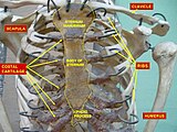
 Thorax. Anterior view.
 -
 Thorax. Anterior view.
 -
 Clearly visible thorax of an artistic gymnast.


_0831.jpg)
See also
References
- ^ "Definition: Thorax". Oxford Learner's Dictionaries.
- ^ "thorax" at Dorland's Medical Dictionary
- ^ Thorax at the U.S. National Library of Medicine Medical Subject Headings (MeSH)
- ^ θώραξ, Henry George Liddell, Robert Scott, A Greek-English Lexicon, on Perseus Digital Library
- ^ "thorax, n.". Oxford English Dictionary. Oxford University Press.
- ^ Shahani, Rohit, MD. (2005). Penetrating Chest Trauma. eMedicine. Retrieved 2005-02-05.
- ^ Chest Diseases Archived 2014-12-16 at the Wayback Machine Retrieved on 2010-1-26
- ^ Atelectasis Lung and Airway Disorders. Retrieved on 2010-1-26
- ^ Pleurisy Lung Diseases. Retrieved on 2010-1-26
External links
- Sam Gon III. "A guide to the Orders of Trilobites". Retrieved August 23, 2005.
| National | |
|---|---|
| Other | |