
Abdominal ultrasonography
| Abdominal ultrasonography | |
|---|---|
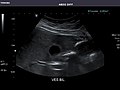
 Medical ultrasound equipment which can be used for abdominal ultrasonography | |
| ICD-9-CM | 88.76 |
| OPS-301 code | 3-059 |
| MedlinePlus | 003777 |

Abdominal ultrasonography (also called abdominal ultrasound imaging or abdominal sonography) is a form of
Abdominal ultrasound examinations are performed by
Medical uses
Abdominal ultrasound can be used to diagnose abnormalities in various internal organs, such as the
Abdominal ultrasound is commonly used in the setting of abdominal pain or an acute abdomen (sudden and/or severe abdominal pain syndrome in which surgical intervention might be necessary), in which it can diagnose appendicitis or cholecystitis.
Ultrasound can also be used if there is suspicion of enlargement of one or more organs, such as used in screening for abdominal aortic aneurysm, investigation for splenomegaly or urinary retention.
Ultrasound can be used for additional anatomical information for patients with an abnormal

It can be used on the abdominal aorta to detect or exclude abdominal aortic aneurysm. For this purpose, the standard aortic measurement for abdominal aortic aneurysm is between the outer margins of the aortic wall.[4]
In cases of infectious mononucleosis, splenomegaly is a common symptom, and health care providers may consider using abdominal ultrasonography to get insight into a person's condition.[5] However, because spleen size varies greatly, ultrasonography is not a valid technique for assessing spleen enlargement and should not be used in typical circumstances or to make routine decisions about fitness for playing sports.[5]
Detecting stones
Ultrasound imaging is useful for detecting stones, for example
Ultrasonography can be used to guide procedures such as treatment for kidney stones with
Liver

In patients with deranged liver function tests, ultrasound may show increased liver size (
Ultrasonography of liver tumors involves two stages: detection and characterization.[citation needed] Tumor detection is based on the performance of the method and should include morphometric information (three axes dimensions, volume) and topographic information (number, location specifying liver segment and lobe/lobes). The specification of these data is important for staging liver tumors and prognosis.[citation needed] Tumor characterization is a complex process based on a sum of criteria leading towards tumor nature definition. Often, other diagnostic procedures, especially interventional ones, are no longer necessary. Tumor characterization using the ultrasound method will be based on the following elements: consistency (solid, liquid, mixed), echogenicity, structure appearance (homogeneous or heterogeneous), delineation from adjacent liver parenchyma (capsular, imprecise), elasticity, posterior acoustic enhancement effect, the relation with neighboring organs or structures (displacement, invasion), vasculature (presence and characteristics on Doppler ultrasonography and contrast-enhanced ultrasound (CEUS).[citation needed]
Renal ultrasonography

Ultrasonography of the kidneys is essential in the diagnosis and management of kidney-related diseases. The kidneys are easily examined, and most pathological changes in the kidneys are distinguishable with ultrasound.[7]
Technique
Advantages of ultrasound imaging of abdominal structures are that the procedure can be performed quickly, bed-side, involves no exposure to
The imaging occurs real-time and without sedation, so that the influence of movements can be assessed quickly. For example, by pressing the ultrasound probe against the gallbladder, a radiological Murphy's sign can be elicited.
Through the abdominal wall, organs inside the
The liver can be imaged by swiping the probe sagittally from medial to lateral at the subcoastal region. However, if majority of the liver parenchyma is located high up in behind the ribs, the subject can be asked to breathe deeply to push down the liver into the abdomen for better visibility of liver. If the liver is still not visualised, then the subject can be rolled to the left lateral position to move the liver out of the ribs. Then, the ultrasound probe is rotated 90 degrees to access the liver in axial plane from the dome of the diaphragm until the lower segment of the liver.[8]
Abdominal Ultrasound (Full Exam)
STRUCTURED REPORT
(Technique: Transabdominal ultrasonography; Device: Toshiba Aplio XG)
Liver: Diffusely homogeneous and normal in echogenicity. No focal mass or contour nodularity. No intrahepatic biliary ductal dilatation.
Portal Vein: Patent main portal vein.
Gallbladder: No stones, wall thickening, or pericholecystic fluid.
Common Bile Duct: Nondilated measuring 1.3 mm at the level of the porta hepatis.
Pancreas: Visualized portions unremarkable.
Spleen: Normal in size.
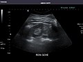
Kidneys: Right and left kidneys measure 11.5 cm and 12 cm in length respectively. No hydronephrosis. Small left lower pole kidney cyst.
Ascites: None.
Aorta: Visualized portions normal in caliber, 16 x 15 mm.
IVC: Normal.
IMPRESSION:
Normal abdominal ultrasound.
-
 Aorta
Aorta -
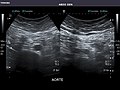 Aorta
Aorta -
 Aorta
Aorta -
 Aorta
Aorta -
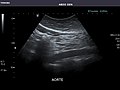 Aorta
Aorta -
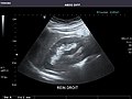 Right kidney
 -
 Right kidney
 -
 Right kidney
 -
 Right kidney
 -
 Right kidney
 -
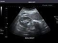 Right kidney
 -
 Right kidney
 -
 Right kidney
 -
 Left kidney
 -
 Left kidney
 -
 Left kidney
 -
 Left kidney
 -
 Left kidney
 -
 Spleen
 -
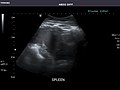 Spleen
 -
 Spleen
 -
 Spleen
 -
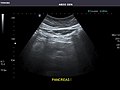 Pancreas
 -
 Pancreas
 -
 Pancreas
 -
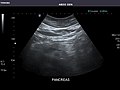 Pancreas
 -
 Pancreas
 -
 Pancreas
 -
 Liver
Liver -
 Liver
Liver -
 Liver
Liver -
 Liver
Liver -
 Liver
Liver -
 Liver
Liver -
 Liver
Liver -
 Liver
Liver -
 Liver
Liver -
 Liver
Liver -
 Liver
Liver -
 Liver
Liver -
 Liver
Liver -
 Liver
Liver -
 Liver
Liver -
 Liver
Liver -
 Liver
Liver -
 Liver
Liver -
 Liver
Liver -
 Gallbladder
 -
 Gallbladder
 -
 Bile duct
 -
 Bile duct
 -
 Gallbladder
 -
 Gallbladder
 -
 Gallbladder
 -
 Gallbladder


See also
References
- ISBN 978-81-312-1574-6. Retrieved 10 April 2011.
- PMID 26171896.
- PMID 23280739.
- ^ a b Jang T (2017-08-28). "Bedside Ultrasonography Evaluation of Abdominal Aortic Aneurysm - Technique". Medscape.
- ^ ABIM Foundation, American Medical Society for Sports Medicine, retrieved 29 July 2014, which cites
- Putukian M, O'Connor FG, Stricker P, McGrew C, Hosey RG, Gordon SM, et al. (July 2008). "Mononucleosis and athletic participation: an evidence-based subject review". Clinical Journal of Sport Medicine. 18 (4): 309–315. S2CID 23780443.
- Spielmann AL, DeLong DM, Kliewer MA (January 2005). "Sonographic evaluation of spleen size in tall healthy athletes". AJR. American Journal of Roentgenology. 184 (1): 45–49. PMID 15615949.
- Putukian M, O'Connor FG, Stricker P, McGrew C, Hosey RG, Gordon SM, et al. (July 2008). "Mononucleosis and athletic participation: an evidence-based subject review". Clinical Journal of Sport Medicine. 18 (4): 309–315.
- ^ Dietrich CF, Tuma J, Badea R (2010-07-28). "Ultrasound of the liver - EFSUMB – European Course Book" (PDF). European federation of societies for ultrasound in medicine and biology (EFSUMB). Archived from the original (PDF) on 2017-08-12. Retrieved 2017-12-22.
- PMID 26838799. (CC-BY 4.0)
- PMID 28191063.
External links
- Abdominal Ultrasound, information for patients from the American College of Radiology and the Radiological Society of North America.
- Abdominal ultrasound from MedlinePlus.
| X-ray/ radiography |
| ||||||||||||
|---|---|---|---|---|---|---|---|---|---|---|---|---|---|
| MRI | |||||||||||||
| Ultrasound | |||||||||||||
| Radionuclide |
| ||||||||||||
| Optical/Laser | |||||||||||||
| Thermography | |||||||||||||
| Target conditions |
| ||||||||||||