Magnaporthe grisea
| Magnaporthe grisea | |
|---|---|

| |
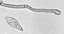
| Conidium and conidiogenous cell | |
| Scientific classification | |
| Domain: | Eukaryota |
| Kingdom: | Fungi |
| Division: | Ascomycota |
| Class: | Sordariomycetes |
| Order: | Magnaporthales |
| Family: | Magnaporthaceae |
| Genus: | Magnaporthe |
| Species: | M. grisea
|
| Binomial name | |
| Magnaporthe grisea (T.T. Hebert) M.E. Barr
| |
| Synonyms | |
|
Ceratosphaeria grisea T.T. Hebert, (1971) | |
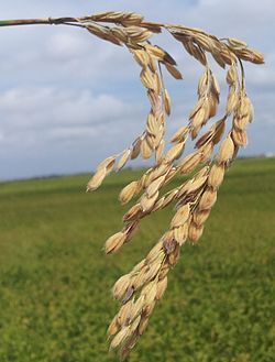
Magnaporthe grisea, also known as rice blast fungus, rice rotten neck, rice seedling blight, blast of rice, oval leaf spot of graminea, pitting disease, ryegrass blast, Johnson spot,
Members of the M. grisea complex can also infect other agriculturally important
Hosts and symptoms

M. grisea is an
Rice blast has been observed on rice strains M-201, M-202, M-204, M-205, M-103, M-104, S-102, L-204, Calmochi-101, with M-201 being the most vulnerable.[16] Initial symptoms are white to gray-green lesions or spots with darker borders produced on all parts of the shoot, while older lesions are elliptical or spindle-shaped and whitish to gray with necrotic borders. Lesions may enlarge and coalesce to kill the entire leaf. Symptoms are observed on all above-ground parts of the plant.[17] Lesions can be seen on the leaf collar, culm, culm nodes, and panicle neck node. Internodal infection of the culm occurs in a banded pattern. Nodal infection causes the culm to break at the infected node (rotten neck).[18] It also affects reproduction by causing the host to produce fewer seeds. This is caused by the disease preventing maturation of the actual grain.[15]
Disease cycle

The pathogen infects as a spore that produces lesions or spots on parts of the rice plant such as the leaf, leaf collar, panicle, culm and culm nodes. Using a structure called an
A single cycle can be completed in about a week under favorable conditions where one lesion can generate up to thousands of spores in a single night. Disease lesions, however, can appear in three to four days after infection.[21] With the ability to continue to produce the spores for over 20 days, rice blast lesions can be devastating to susceptible rice crops.[22]
Infection of
Environment
Rice blast is a significant problem in temperate regions and can be found in areas such as irrigated lowland and upland.
In terms of control, excessive use of
Geographical distribution
Wheat blast was found in the 2017-2018 rainy season in Zambia, in the Mpika district of the Muchinga Province.[25][26]
In February 2016 a devastating wheat epidemic struck Bangladesh.[27][28] Transcriptome analysis showed this to be an M. grisea lineage most likely from Minas Gerais, São Paulo, Brasília, and Goiás states of Brazil and not from any geographically proximate strains.[27][28] This successful diagnosis shows the ability of genetic surveillance to untangle the novel biosecurity implications of transcontinental transportation[27][28] and allows the Brazilian experience to be rapidly applied to the Bangladeshi situation.[27][28] To that end the government has set up an early warning system to track its spread through the country.[28]
Management

This fungus faces both
: 107–108The wheat blast strain can be diagnosed by sequencing.
Some innovative biologically-imitative fungicides are being developed from
Importance
Rice blast is the most important disease concerning rice crops in the world. Since rice is an important food source for much of the world, its effects have a broad range. It has been found in over 85 countries across the world and reached the United States in 1996. Every year the amount of crops lost to rice blast could feed 60 million people. Although there are some resistant strains of rice, the disease persists wherever rice is grown. The disease has never been eradicated from a region.[32]
Strains
Three strains, albino (defined by a mutation at the ALB1 locus), buff (BUF1), and rosy (RSY1) have been extensively studied because they are nonpathogenic. This has been found to be due to nonuse of melanin, which is a virulence factor in M. grisea.[1]: 184 The pathovar triticum strain (M. o. pv. triticum) causes the wheat blast disease.[12]
Genetics
A
Because signal links between MAPKs and
The transaminase alanine: glyoxylate aminotransferase 1 (AGT1) has been shown to be crucial to the pathogenicity of M. grisea through its maintenance of redox homeostasis in peroxisomes. Lipids transported to the appressoria during host penetration are degraded within a large central vacuole, a process that produces fatty acids. β-Oxidation of fatty acids is an energy producing process that generates Acetyl-CoA and the reduced molecules FADH2 and NADH, which must be oxidized in order to maintain redox homeostasis in appressoria. AGT1 promotes lactate fermentation, oxidizing NADH/FADH2 in the process.[33]
M. grisea mutants lacking the AGT1 gene were observed to be nonpathogenic through their inability to penetrate host surface membranes. This indicates the possibility of impaired lipid utilization in M. grisea appressoria in the absence of the AGT1 gene.[34]
Biochemistry of host-pathogen interactions
A 2010 review reported
See also
- Corn grey leaf spot, a similar disease in maize/corn
- Gray leaf spot, a similar disease in other grasses
References
- ^ PMID 14527276.
Three mutants of M. grisea, albino, buff, and rosy (corresponding to the ALB1, BUF1, and RSY1 loci, respectively), have been studied extensively and are nonpathogenic. This is due to an inability to cross the plant cuticle because of the lack of melanin deposition in the appressorium.
- Centre for Agriculture and Bioscience International.
- S2CID 42684382.
- S2CID 549194.
- PMID 15846337.
- PMID 21156541.
- ^ Magnaporthe grisea Archived 2007-10-12 at the Wayback Machine at Crop Protection Compendium Archived 2007-07-16 at the Wayback Machine, CAB International
- ISSN 1935-9411.
- PMID 25444707.
- S2CID 45126761.
- S2CID 14478689.
- ^ S2CID 235049332.
- ^ S2CID 20430256.
- ^ PMID 15802503.
- ^ University of California-Davis (UCD). Archived from the originalon 2006-09-11. Retrieved 2014-02-25.
- University of California Integrated Pest Management(UC-IPM)
- ^ Rice Blast at the Online Information Service for Non-Chemical Pest Management in the Tropics
- ^ Rice Blast Archived 2010-10-20 at the Wayback Machine at Factsheets on Chemical and Biological Warfare Agents
- PMID 29567712.
- Elsevier Academic Press.
- S2CID 42684382.
- ^ Diagnostic Methods for Rice Blast[permanent dead link] at PaDIL Plant Biosecurity Toolbox
- ^ a b
Liu, Xinyu; Zhang, Zhengguang (2022). "A double-edged sword: reactive oxygen species (ROS) during the rice blast fungus and host interaction". The FEBS Journal. 289 (18). S2CID 237340135.
- ^ a b Kuyek, Devlin (2000). "Implications of corporate strategies on rice research in asia". Grain. Retrieved 2010-10-20.
- ^ "Researchers in Zambia confirm: Wheat blast has made the intercontinental jump to Africa". 24 September 2020. Archived from the original on 6 October 2021. Retrieved 1 October 2020.
- S2CID 221843315.
- ^ PMID 27716181.
- ^ a b c d e "New infographic highlights an early warning system for wheat blast in Bangladesh". CGIAR WHEAT. 2020-07-15. Archived from the original on 2020-12-01. Retrieved 2020-12-26.
- ^ Kurahasi, Yoshio (1997). "Biological Activity of Carpropamid (KTU 3616): A new fungicide for rice blast disease". Journal of Pesticide Science. Retrieved 2014-02-25.
- S2CID 199492358.
- ^ S2CID 237433001.
- ^ Rice Blast Archived 2010-07-31 at the Wayback Machine at Cereal Knowledge Bank
- PMID 22558413.
- PMID 22899049.
- PMID 19400646.
- ^ PMID 34379774.
- ^ PMID 35635051.
- S2CID 29633821.
Further reading
- , February 11, 1998
- CIMMYT. What is wheat blast?, 2019.
- Kadlec, RP. Biological Weapons for Waging Economic Warfare, Air & Space Power Chronicles
- NSF. Microbial Genome Helps Blast Devastating Rice Disease, April 21, 2005
- United States Congress. Testimony of Dr. Kenneth Alibek, 1999
- "What is wheat blast?". CIMMYT. 2019-12-11. Retrieved 2020-12-21.
- "What is wheat blast? How can I manage it?" (PDF). CIMMYT.
- 加藤 Katō, 肇 Hajime; 山口 Yamaguchi, 富夫 Tomio; 西原 Nishihara, 夏樹 Natsuki (1976). "The perfect state of Pyricularia oryzae Cav. in culture". ISSN 1882-0484.
- "Blast (node and neck)". Rice Knowledge Bank. IRRI (International Rice Research Institute). 2017-08-15. Retrieved 2021-03-04.
